 |
Case Report
The abdominal cocoon: A management dilemma
1 Surgical registrar, Department of General Surgery, Peninsula Health PO Box 52, 2 Hastings Road, Frankston, Victoria, Australia
2 Senior Surgical Registrar, Department of General Surgery
3 Royal Darwin Hospital, Northern Territory, Australia
4 Peter macCullum hospital, Melbourne, Australia
5 Monash Health, Melbourne, Australia
Address correspondence to:
Amin Tanveer
Department of General Surgery, Peninsula Health PO Box 52, 2 Hastings Road, Frankston, Victoria, Australia, Frankston, Melbourne, Victoria 3199,
Australia
Message to Corresponding Author
Article ID: 100053Z06AT2018
Access full text article on other devices

Access PDF of article on other devices

How to cite this article
Tanveer A, Arachchi A, Kalgutkar S, Lynch C, Suhardja T. The abdominal cocoon: A management dilemma. Case Rep Int 2018;7:100053Z06AT2018.ABSTRACT
Abdominal cocoon is a rare entity with no known aetiology and is a clinical curiosity in the surgical field, and a management dilemma for surgeons. Treatment may include excision of the accessory peritoneal sac with lysis of the inter-loop adhesions. Bowel resection is unnecessary unless a nonviable segment is found. However, if there are no signs of acute abdominal symptoms after diagnosing abdominal cocoon syndrome pre-operatively, surgery is unnecessary. We presenting a case of a 47-year-old male who had laparotomy noted an entirely frozen abdomen which was depicted as a cocoon. The stomach was only identified due to feeling the position of the NGT, no other anatomy was delineated. A peritoneal biopsy was taken prior to closure. Post operatively he was given parenteral and trickle enteral nutritional support and was discharged home with PEG gastrostomy for feeding.
Keywords: Abdominal cocoon, Idiopathic encapsulating peritonitis, Sclerosing encapsulating peritonitis
INTRODUCTION
Abdominal cocoon is a rare entity with no known aetiology and is a clinical curiosity in the surgical field. It is characterized by encapsulation of part or whole of the small bowel by a fibrous membrane [1] and can present with intestinal obstruction [2].
Abdominal cocoon phenomenon is a rare case of sclerosing encapsulating peritonitis of the abdomen; first described in 1970s. Varying lengths of small bowel are encased in a fibrocollagenous sac; and since its description there has been various amendments to its understanding and terminology [3],[4],[5].
Abdominal cocoon remains a management dilemma for surgeons. Treatment may include excision of the accessory peritoneal sac with lysis of the inter-loop adhesions. Bowel resection is unnecessary unless a nonviable segment is found. However, if there are no signs of acute abdominal symptoms after diagnosing abdominal cocoon syndrome pre-operatively, surgery is unnecessary.
CASE REPORT
A 47-year-old indigenous male presented to our institution with abdominal pain. Clinical and radiological features were suggestive of high grade gastric outlet obstruction.
He has a background of rate controlled atrial fibrillation and Cor Pulmonale secondary to Obstructive Sleep Apnoea.
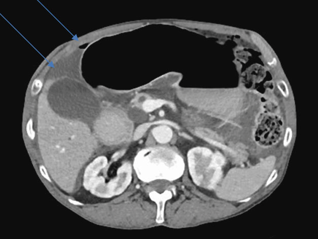
On presentation, an abdominal computed tomography (CT) contrast study noted the stomach; first, second and proximal third parts of the duodenum to be markedly distended, with a transition point at the mid third part of duodenum with no mass or thickening identified.
An emergency gastroscopy reported a grossly dilated stomach to second part of duodenum with difficulty passing the scope due to perhaps an intraluminal obstruction. A subsequent laparotomy noted an entirely frozen abdomen which was depicted as a cocoon (Figures 1, 2). The stomach was only identified due to feeling the position of the NGT, no other anatomy was delineated. A peritoneal biopsy was taken prior to closure, which was reported a reactive myofibroblastic proliferation. Histopathological examination revealed spindle cell proliferation (Figure 3). Post operatively, he was given parenteral and trickle enteral nutritional support, and was discharged home with PEG gastrostomy for feeding. Unfortunately, patient did not attend hospital outpatient clinic for follow-up.
DISCUSSION
Abdominal cocoon phenomenon is a rare case of sclerosing encapsulating peritonitis of the abdomen; first described in 1970s. Varying lengths of small bowel are encased in a fibrocollagenous sac; and since its description there has been various amendments to its understanding and terminology [3],[4],[5].
Type I abdominal cocoon is partial encapsulation of the intestine, Type II is complete encapsulation of the entire intestine, and Type III cocoon syndrome is encasement of the entire intestine along with other intraabdominal organs like appendix, caecum, ascending colon and ovaries. Encapsulation of bowel leads to acute or chronic bowel obstruction [3],[5],[6].
Abdominal cocoon has been classified as primary and secondary based on whether it is idiopathic or has a definite cause. The aetiology of the primary form is uncertain, while secondary abdominal cocoon is thought to be secondary to tuberculosis, desmoid tumors and an association with Familial Adenomatous Polyposis (FAP). Complications from long term peritoneal dialysis and porto-venous shunting have been described [3],[7],[8].
In our case the peritoneal biopsies were reported as a spindle cell proliferation; with negative staining for carcinoid, GIST and other tumors and thus inconclusive in terms of management. An annotation of the initial histopathology reported a reactive myofibroblastic proliferation and no features of a diagnostic malignant mesothelioma.
Options for management of this 47-year-old gentleman were irresolute in terms of further surgery and nutrition. Formal advice and suggestions for management were sought from tertiary health care institutions in Australia. However, if there are no signs of acute abdominal symptoms after diagnosing abdominal cocoon syndrome pre-operatively, surgery is unnecessary [9].
Suggestions included venting PEG gastrotomy for feeding – with radiological guided feeding distal to the small bowel stricture for short term feeding. Tamoxifen and non-steroidals were also recommended as a more conservative approach to what is, a benign process. Based on the macroscopic appearance of the small bowel, it was suggested his long term prognosis to be likely poor.
We are thus left with a challenging entity of the abdominal cocoon; and perhaps with unanswered questions in terms of future treatment goals. However, if there are no signs of acute abdominal symptoms after diagnosing abdominal cocoon syndrome pre-operatively, surgery is unnecessary [9].
CONCLUSION
Abdominal cocoon remains a management dilemma for surgeons. Treatment may include excision of the accessory peritoneal sac with lysis of the inter-loop adhesions. Bowel resection is unnecessary unless a nonviable segment is found.
REFERENCE
1.
Kaushik R, Punia RP, Mohan H, Attri AK. Tuberculous abdominal cocoon – a report of 6 cases and review of the Literature. World J Emerg Surg 2006 Jun 27;1:18. [CrossRef]
[Pubmed]

2.
Lalloo S, Krishna D, Maharajh J. Case report: Abdominal cocoon associated with tuberculous pelvic inflammatory disease. Br J Radiol 2002 Feb;75(890):174–6. [CrossRef]
[Pubmed]

3.
Sharma D, Nair RP, Dani T, Shetty P. Abdominal cocoon-A rare cause of intestinal obstruction. Int J Surg Case Rep 2013;4(11):955– 7. [CrossRef]
[Pubmed]

4.
Singh B, Gupta S. Abdominal cocoon: A case series. Int J Surg 2013;11(4):325–8. [CrossRef]
[Pubmed]

5.
Mbanje C, Mazingi D, Forrester J, Mungazi SG. Peritoneal encapsulation syndrome: A case report and literature review. Int J Surg Case Rep 2017 Nov 9;41:520–3. [CrossRef]
[Pubmed]

6.
Jagdale A, Prasla S, Mittal S. Abdominal cocoon - A rare etiology of intestinal obstruction. J Family Med Prim Care 2017 Jul–Sep;6(3):674–6. [CrossRef]
[Pubmed]

7.
Ghazan-Shahi S, Bargman JM. Case report: Acute bowel obstruction with an isolated transition point in peritoneal dialysis patients; a presentation of encapsulating peritoneal sclerosis? BMC Nephrol 2016 Jan 5;17:1. [CrossRef]
[Pubmed]

8.
Tannoury JN, Abboud BN. Idiopathic sclerosing encapsulating peritonitis: Abdominal cocoon. World J Gastroenterol 2012 May 7;18(17):1999–2004. [CrossRef]
[Pubmed]

9.
Solmaz A, Tokoçin M, Arici S, et al. Abdominal cocoon syndrome is a rare cause of mechanical intestinal obstructions: A report of two cases. Am J Case Rep 2015 Feb 11;16:77–80. [CrossRef]
[Pubmed]

SUPPORTING INFORMATION
Author Contributions
Amin Tanveer - Substantial contributions to conception and design, Acquisition of data, Analysis of data, Interpretation of data, Drafting the article, Revising it critically for important intellectual content, Final approval of the version to be published
Asiri Arachchi - Analysis of data, Interpretation of data, Final approval of the version to be published
Sanjay Kalgutkar - Analysis of data, Interpretation of data, Revising it critically for important intellectual content, Final approval of the version to be published
Craig Lynch - Analysis of data, Interpretation of data, Revising it critically for important intellectual content, Final approval of the version to be published
Thomas Suhardja - Analysis of data, Interpretation of data, Drafting the article, Revising it critically for important intellectual content, Final approval of the version to be published
Guarantor of SubmissionThe corresponding author is the guarantor of submission.
Source of SupportNone
Consent StatementWritten informed consent was obtained from the patient for publication of this case report.
Data AvailabilityAll relevant data are within the paper and its Supporting Information files.
Conflict of InterestAuthors declare no conflict of interest.
Copyright© 2018 Amin Tanveer et al. This article is distributed under the terms of Creative Commons Attribution License which permits unrestricted use, distribution and reproduction in any medium provided the original author(s) and original publisher are properly credited. Please see the copyright policy on the journal website for more information.


