|
Case Report
Mayer Rokitansky Küster Hauser Syndrome and Endometriosis (MRKH): A case report
1 Residency Program, Department of Gynecology and Obstetrics, Federal University of Amazon – School of Medicine, UFAM – Manaus, Amazonas, Brazil
2 Intern at School of Medicine, Federal University of Amazon, UFAM – Manaus, Amazonas, Brazil
3 Faculty member, Department of Gynecology and Obstetrics, Federal University of Amazon – School of Medicine, UFAM – Manaus, Amazonas, Brazil
Address correspondence to:
Isabela Goncalves Feitosa
Intern at School of Medicine, Federal University of Amazon, UFAM, Manaus, Amazon, Brazil – Manaus, Amazonas,
Brazil
Message to Corresponding Author
Article ID: 100056Z06FS2019
Access full text article on other devices

Access PDF of article on other devices


How to cite this article
da Silva FGN, Bulcão FEA, Feitosa IG, dos Santos ES, da Silva MRV, da Silva PRL. Mayer Rokitansky Küster Hauser Syndrome and Endometriosis (MRKH): A case report. Case Rep Int 2019;8:100056Z06FS2019.ABSTRACT
Introduction: MRKH is a rare congenital malformation that affects about 1:4500 female newborns. Its cause is unknown, but the reproductive abnormalities are due to lack of development of the Müllerian ducts between the fifth and the sixth weeks of gestation. This syndrome is characterized by complete or partial vaginal agenesis, tubal, and uterine-cervical abnormalities. In clinical presentation, the disease typically presents itself as primary amenorrhea in an adolescent who has secondary sexual characteristics compatible with their age and may be accompanied by cyclic dysmenorrhea when a rudimentary uterus with functional endometrium is present.
Case Report: This case report describes follow-up appointments and outcomes of a young female patient who looked for gynecological care at The Getúlio Vargas University Hospital. She presented primary amenorrhea with normal development of secondary sexual characteristics. Within the diagnostic investigation it was demonstrated that there was MRKH and endometriosis occurrence. This specific patient had progressive cyclic pelvic pain that didn’t respond to conservator treatment. Therefore, laparoscopic hysterectomy was performed.
Conclusion: Diagnosis is most often based on a clinical suspicion associated with a detailed physical and gynecological examination. The anatomical treatment of the syndrome is surgical. The frequent association of this disease with somatic and psychosocial disorders requires a multidisciplinary therapeutic.
Keywords: Abnormalities; Amenorrhea, Endometriosis, Paramesonephric (Müllerian) ducts
INTRODUCTION
The Mullerian agenesis or Mayer Rokitansky Küster Hauser (MRKH) syndrome is identified by vaginal atresia, uterus, and tubal abnormalities that can be presented as hypoplasia or the absence of those structures [1]. The uterus remnants may or may not contain functioning endometrial tissue and the vagina can consist of a small dimple between the labia majora, with a length up to 2 and a half inches long (6 cm) [2]. However, most MRKH patients present secondary sexual characteristics such as external vagina appearance, breast growth, and pubic hair once the ovaries are typically functioning and existent [1],[3].
There is a possibility the genital defect of the MRKH syndrome occurs due to the disruption or dysregulation of the fusion and canalization processes of the Mullerian ducts. Carrol et al., in 2005, demonstrated that heterozygotic mutations on Wn9tb gene can be related to the syndrome’s etiology [4],[5].
Primary amenorrhea is the typical clinical presentation (58%), cyclic abdominal or pelvic pain and dyspareunia (14%) may occur in teenage patients with secondary sexual characteristics compatible with their age [1]. This malformation affects 1 per 4,000-5,000 female newborn children and is the second most frequent cause of primary amenorrhea, after gonadal dysgenesis [6],[7],[8],[9]. Most of the cases appear as a sporadic occurrence, but there are family related cases described in the literature [10],[11]. The most common karyotype is 46, XX [9].
MRKH is classified in two ways according to the structural or reproductive system impairment. Type I (Isolated) is the typical syndrome – there are restrict alterations to the reproductive system such as vaginal agenesis, symmetrical uterus remnants, fallopian, and typical ovaries [7]. Type II or MURCS association (Mullerian duct aplasia, Renal dysplasia, and Cervical Somite anomalies), is the atypical presentation, there is asymmetry of the uterus remnants and tubal abnormalities. The second type is associated with ovary diseases, kidney, bone, hearing, or other associated congenital malfunctions [1],[8]. It corresponds to the severe form of this syndrome. The American Society for Reproductive Medicine classifies hypoplasia/ agenesis into 5 subtypes: vaginal, cervical, fundal, tubal and combined aplasia [12].
The rudimentary uterus present in MKRH may still have functioning endometrium tissue [13]. Although, it is not commonly described, pelvic pain in those women diagnosed with MKRH syndrome may indicate the presence of functioning endometrium, resulting in uterine dilatation. It resembles the pain observed in women diagnosed with other Mullerian obstructive abnormalities. Also, the retrograde flow of the menstrual fluid may result in peritoneal irritation and extrauterine endometrial tissue implantation. Endometriosis is something that has been often reported by women with MRKH [12]. Endometriosis is histologically characterized by the ectopic growth of endometrium-like tissue in response to cyclic hormonal action [14], it is linked to pain during menses or intercourse.
Another theory to explain the presence of endometriosis in those patients would be celomic metaplasia. In such cases, the endometriosis might develop through metaplasia resulted by aberrant activation of genes on the peritoneum that are usually active during embryonic development of the female genital tract, including the endometrial gland development [14].
This case report presents the case of a young woman who looked for gynecological care presenting amenorrhea, moderate to severe cyclic low abdominal pain with secondary sexual characteristics compatible with her age.
CASE REPORT
A nonsexually active, 19-year-old female, born in the state of Amazonas, Brazil, presented herself in 2001 at the Obstetrics and Gynecology Clinic of the Getúlio Vargas University Hospital. Her follow-up was due to primary amenorrhea associated to cyclic lower abdominal pain. At the moment, she had not expressed any symptomology. During the physical examination, it was observed that the patient had normal secondary sexual characteristics corresponding to her chronological age.
The gynecological examination revealed a eutrophic vulva, labia majora, and labia minora which showed no alteration, and the hymen was intact. For that reason, no specular examination was performed, and a swab was used to measure the length of the vaginal canal (3 cm long). The patient brought a negative Progesterone and Estrogen test and pelvic ultrasound that evidenced regular uterus and ovaries. The karyotype analysis and another ultrasonography was done to assess the internal genitalia. Karyotype was 46, XX. Ultrasonography didn’t reveal significant changes.
The patient was absent from the clinic for almost two years and returned due to the increase of abdominal pain. She was prescribed oral contraceptive pills (OCPs) and a new imaging examination was done. The abdominal ultrasonography exhibited a 16,8 cm³ uterus and atrophic cervix. At the follow-up appointment, patient complaints were still related to abdominal cyclic pain that would last up to five days and not cease to the use of over the counter analgesics. It was oriented to the patient continued use of the oral contraceptive pills, NSAIDs, and pelvic sonogram.
Five months later, there had been persistence of pain in the pelvic area. Hence, the patient had started sexual intercourse, it was possible to perform the speculum examination, which demonstrated a blind-ended vagina and absence of the cervix (Figure 1). The patient was once again oriented to maintain the OCPs and to return to follow up appointments every three months.
The patient denied the use of OCPs and she also referred reoccurrence of the cyclic lower abdominal pain. New transvaginal sonography revealed the presence of a moderate amount of free fluid in the pelvis and a small thick fluid filled cyst on the right ovary (endometrioma?). At this point, OCPs were suspended and medroxyprogesterone Acetate (MPA) 150 mg quarterly was initiated resulting in the patient’s pain complaints improvement.
After seven years she regresses to the clinic bringing a –2.9 standard deviation bone mineral density test result. MPA was suspended, oral contraceptive pills restarted and Sodium Alendronate prescribed concomitantly. Even so, due to the abdominal pain persistence and thinking of the clinical findings that resembled an endometrioma, it was decided to switch the OCPs to Dienogest.
In 2014, patient was submitted to a video laparoscopy. Its report suggested the duplicity of a small rudimentary left sided uterus. Presence of cyst-like formation on the right ovary topography displaying purple lesions on its surface and surroundings, uterus and bladder. Right fallopian tube increased in size and significant congestion. Right salpingectomy and right ovarian cyst removal were performed and both surgical pieces were sent to pathology for analysis. The result of the histopathological report was a serous cystadenoma and tubal congestion.
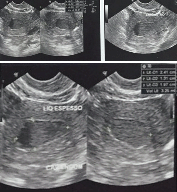
A year after the procedure patient returned with daily pelvic pain complaints not related to physical effort with vomit depending on the degree of pain. She was continuously using OCPs prescribed by different clinic. A new sonogram revealed a 2.4x1.7 cm hypoechoic imaging in the uterus cavity suggesting hematometra and a 3.1x1.9x2.3 cm solid-cystic imaging on left ovary (Figure 2). Thus, a possibility of hysterectomy was indicated.
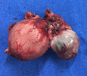
In 2016, laparoscopic hysterectomy and left salpingooophorectomy were performed. The uterus was bicornuate, the right side was rudimentary with typical ovary and the left side showed an increase in size and voluminous endometrioma on left ovary (Figure 3). The vagina ended in a blind pouch and there was agenesis of the uterus cervix – which was also reported in the histopathological analysis.
The patient is currently asymptomatic and is not using any medication. Laboratory blood work and new imaging sonograms did not reveal any evident alterations. She was requested a new Bone Mineral Density test in September 2018 to evaluate the osteopenia progress. The patient still awaits availability to schedule the examination through the Public Health System of Brazil.

DISCUSSION
Mayer Rokitansky Küster Hauser (MRKH) syndrome is a fertility obstacle for young adult women. These patients usually manifest psychological consequences – such as anxiety, lower self-esteem, and lower life quality – associated with this diagnose [15],[16],[17]. As the main outcome of the MRKH syndrome includes partial or complete vaginal agenesis, uterine and cervical abnormalities – for instance, the cervix aplasia/ hypoplasia – primary amenorrhea is the main reason women go to find medical assistance [7],[18],[19].
Secondary to amenorrhea, other symptoms may be present at the Mullerian malformations and are associated with severe health conditions that might implicate serious consequences to the patient’s health. For example, the obstructive abnormalities on which the uterus has a functioning endometrium but no communication to the vaginal canal, or it’s associated to aplasia or dysplasia of the cervix and/or the vagina. Therefore, it can present itself along with cyclic lower abdominal pain, endometriosis and dyspareunia [20].
The clinical suspicion diagnosis starts when the patient is a 14 to 18 years old woman with a normal female phenotype and has physiological development of the secondary sexual characteristics, yet without menstruation periods. Those with Mullerian remnants may develop chronic cyclic pelvic pain [19],[21]. Just as the patient presented in this case report, the secondary sexual characteristics and external genitalia are usually normal, and so are the hormonal levels; a shortened cuneiform vagina ending on a blind pouch and intact hymen are typically present [22].
The diagnosis is done by the high clinical suspicion correlated to a thorough physical and gynecological examination. In 2011, it was demonstrated that the clinical examination and ultrasonography were enough to correctly classify the syndrome. The efficacy was similar to the magnetic resonance imaging alone, but before puberty, the sonogram of those patients may be difficult to interpret and be misleading [6],[22].
Exploratory laparoscopy is not mandatory, but it’s considered to be the gold standard when it comes to the diagnose and it’s important for the evaluation and management of the patients who report pelvic pain. Laparoscopy is indicated for MRKH syndrome when the imaging exams and/or hysteroscopy are unsatisfactory [23],[24],[25].
At the case report presented there is an association of cervical aplasia and uterovaginal hypoplasia of the MRKH syndrome in association to endometriosis. Endometriosis is the cause of dysmenorrhea for 20% of women who present any sort of Mullerian abnormality [12].
The treatment is based on medical therapy, which includes hormonal and non-hormonal medication, such as analgesics and surgical procedures aiming at efficient pain management [22]. The need for surgical correction of uterine malformation varies according to each case and depends on the signs and symptoms presented, besides the uterus anatomy and patient’s history [20].
The anatomical correction of the syndrome is creating a new vaginal canal through surgical or non-surgical procedures in order to allow the patient to perform sexual activities. However, vaginal elongation should wait until the patient has been oriented and feels secure and emotionally stable about the procedure and expresses the desire to proceed.
Surgical procedures with removal of the uterine remnants also aim to avoid endometriosis development, similar to the case reported in this article [10],[18],[26]. Due to the pain associated with the obstruction of the structures, if endometrium tissue is identified by MRI, it’s indicated to have all the uterine remnants surgically removed. Bilateral removal is a possibility even if functioning endometrium is only found in one of the uterine horns [12],[27].
MRKH syndrome diagnostic brings a significant psychological impact on the patients’ lives and is related to depression, low self-esteem and personality disorders – factors which directly affect the patient’s life quality [16],[17],[28].
This report displays a case that did not respond to conservative treatment, once the low abdominal pain was progressive. Therefore, in order to promote a better quality of life to the patient, it was performed laparoscopic surgery – which confirmed the diagnose and removed the uterine remnants. The patient was consulted on a possible vaginal reconstruction surgery but refused due to reporting a satisfactory sexual life.
CONCLUSION
Mayer Rokitansky Küster Hauser (MRKH) syndrome may be a rare anomaly of the Mullerian duct but it has a deep effect on the life of those patients affected by it. Not only because of genetic malformation but because they come to find themselves unable to biologically conceive. Most of the patients need vaginal reconstruction surgery in order to perform vaginal penetration intercourse and some even start to question their own female identity. It’s imperative to ensure the patient receives a multiprofessional assessment, from psychological counseling – including some of the patient’s close relatives as well – up to surgical procedures. The patient should also be screened for other congenital malformations, be oriented about her diagnose and maintain a gynecological care routine as any other woman without Mullerian agenesis. Moreover, we must always consider the social and epidemiological background of the patients. A patient who has been attended by the Public Health System of Brazil in comparison to other parts of the world add multi-dynamic issues regarding the adequate access and management of the patient’s treatment and medical monitoring.
REFERENCE
1.
Rezende DF, Filho AVR, Oliveira GN, Mourão HA, Neto JMP, Sampaio PRL. Síndrome de Mayer-Rokitansky-Küster-Hauser: Uma revisão da literatura. Rev Med Saude Brasilia 2013;2(2):90–9.

2.
Nodale C, Ceccarelli S, Giuliano M, et al. Gene expression profile of patients with Mayer-Rokitansky-Küster-Hauser syndrome: New insights into the potential role of developmental pathways. PLoS one 2014;9(3):e91010. [CrossRef]
[Pubmed]
3.
Oppelt PG, Lermann J, Strick R, et al. Malformations in a cohort of 284 women with Mayer-Rokitansky-Küster-Hauser syndrome (MRKH). Reprod Biol Endocrinol 2012;10:57. [CrossRef]
[Pubmed]
4.
Bousfiha N, Errarhay S, Saadi H, Ouldim K, Bouchikhi C, Banani A. Gonadal dysgenesis 46, XX associated with Mayer-Rokitansky-Kuster-Hauser syndrome: One case report. Obstet Gynecol Int 2010;2010:847370. [CrossRef]
[Pubmed]
5.
Wang M, Li Y, Ma W, et al. Analysis of WNT9B mutations in Chinese women with Mayer-Rokitansk-Küster-Hauser syndrome. Reprod Biomed online 2014;28(1):80–5. [CrossRef]
[Pubmed]
6.
Lermann J, Mueller A, Wiesinger E, et al. Comparison of different diagnostic procedures for the staging of malformations associated with MayerRokitansky-Küster-Hauser syndrome. Fertil Steril 2011;96(1):156–9. [CrossRef]
[Pubmed]
7.
Gutsche RM, Chagas LA, Leal R, Cunha AL, Djahjah MCR. Síndrome de Mayer-Rokitansky-Kuster-Hauser: Relato de caso e revisão da literatura. Radiol Bras 2011;44(3):192–4 [CrossRef]
8.
Barbosa G, Guzmán MV. Adolescente com Síndrome de Mayer-Von Rokitansky-Küster-Hauser: La importancia de un manejo integral multidisciplinario. Revista Colombiana de Obstetricia y Ginecología 2006;57(4):305.
9.
Bombard DS 2nd, Mousa SA. Mayer-Rokitansky-Kuster-Hauser syndrome: Complications, diagnosis and possible treatment options: A review. Gynecol Endocrinol 2014;30(9):618–23. [CrossRef]
[Pubmed]
10.
Sousa S, Silva C, Rodrigues C, Leite H, Geraldes F, Àguas F. Abordagem diagnóstica e terapêutica no síndrome de Mayer-Rokitansky-Küster-Hauser. Acta Obstet Ginecol Port 2014;8(1):8–13.
11.
Guerrier D, Mouchel T, Pasquier L, Pellerin I. The Mayer-Rokitansky-Küster–Hauser syndrome (congenital absence of uterus and vagina) – phenotypic manifestations and genetic approaches. J Negat Results Biomed 2006;5:1. [CrossRef]
[Pubmed]
12.
Marsh CA, Will MA, Smorgick N, Quint EH, Hussain H, Smith YR. Uterine remnants and pelvic pain in females with Mayer-Rokitansky-Küster-Hauser syndrome. J Pediatr Adolesc Gynecol 2013;26(3):199–202. [CrossRef]
[Pubmed]
13.
Escobar ME, Gryngarten M, Del Rey G, et al. Síndrome de Rokitansky (agenesia úterovaginal): Aspectos clínicos, diagnósticos y terapéuticos. Arch Argent Pediatr 2007;105(1):25–31.
14.
Klemmt PAB, Starzinski-Powitz A. Molecular and cellular pathogenesis of endometriosis. Curr Womens Health Rev 2018;14(2):106–16. [CrossRef]
[Pubmed]
15.
Reichman DE, Laufer MR. Mayer-Rokitansky-KüsterHauser syndrome: Fertility counseling and treatment. Fertil Steril 2010;94(5):1941–3. [CrossRef]
[Pubmed]
16.
Bean EJ, Mazur T, Robinson AD. Mayer-Rokitansky-Küster-Hauser syndrome: Sexuality, psychological effects, and quality of life. J Pediatr Adolesc Gynecol 2009;22(6):339–46. [CrossRef]
[Pubmed]
17.
Sanfilippo JS. Mayer-Rokitansky-Kuster-Hauser (MRKH) syndrome: It’s more than the anatomy. J Pediatr Adolesc Gynecol 2009;22(6):337–8. [CrossRef]
[Pubmed]
18.
Londra L, Chuong FS, Kolp L. Mayer-RokitanskyKuster-Hauser syndrome: A review. Int J Womens Health 2015;7:865–70. [CrossRef]
[Pubmed]
19.
Patnaik SS, Brazile B, Dandolu V, Ryan PL, Liao J. Mayer-Rokitansky-Küster-Hauser (MRKH) syndrome: A historical perspective. Gene 2015;555(1):33–40. [CrossRef]
[Pubmed]
20.
Committee on adolescent health care. ACOG committee opinion no. 728: Müllerian agenesis: Diagnosis, management, and treatment. Obstet Gynecol 2018;131(1):e35–e42. [CrossRef]
[Pubmed]
21.
Preibsch H, Rall K, Wietek BM, et al. Clinical value of magnetic resonance imaging in patients with MayerRokitansky-Küster-Hauser (MRKH) syndrome: Diagnosis of associated malformations, uterine rudiments, and intrauterine endometrium. Eur Radiol 2014;24(7):1621–7. [CrossRef]
[Pubmed]
22.
Troncon JK, Zani AC, Vieira AD, Poli-Neto OB, Nogueira AA, Rosa-E-Silva JC. Endometriosis in a Patient with Mayer-Rokitansky-KüsterHauser syndrome. Case Rep Obstet Gynecol 2014;2014:376231. [CrossRef]
[Pubmed]
23.
Pizzo A, Laganà AS, Sturlese E, et al. MayerRokitansky-Kuster-Hauser syndrome: Embryology, genetics, and clinical and surgical treatment. ISRN Obstet Gynecol 2013;2013:628717. [CrossRef]
[Pubmed]
24.
Antunes MRP. Malformações uterinas - do diagnóstico ao tratamento. Artigo de Revisão 2016;24–34.
25.
Ávila-Vergara MA, León-Álvarez DA, López-Villegas MN, Quintero-Medrano SM, Angulo-Bueno GF, Vadillo-Ortega F. Mayer-Rokitansky-Küster-Hauser syndrome: Two cases report. [Article in Spanish]. Ginecol Obstet Mex 2015;83(3):199–205.
[Pubmed]
26.
Morcel K, Camborieux L; Programme de recherches sur les aplasies müllériennes, Guerrier D. MayerRokitansky-Küster-Hauser (MRKH) syndrome. Orphanet J Rare Dis 2007;2:13. [CrossRef]
[Pubmed]
27.
Cho MK, Kim CH, Oh ST. Endometriosis in a patient with Rokitansky-Kuster-Hauser syndrome. J Obstet Gynaecol Res 2009;35(5):994–6. [CrossRef]
[Pubmed]
28.
Elliott JE, Abduljabar H, Morris M. Presurgical management of dysmenorrheal and endometriosis in a patient with Mayer-Rokitansky-Kuster-Hauser syndrome. Fertil Steril 2011;96(2)e86–9. [CrossRef]
[Pubmed]
SUPPORTING INFORMATION
Author Contributions
Fernanda Goulart Nogueira da - Substantial contributions to conception and design, Acquisition of data, Drafting the article, Revising it critically for important intellectual content, Final approval of the version to be published
Felipe Everton Araújo Bulcão - Substantial contributions to conception and design, Acquisition of data, Drafting the article, Revising it critically for important intellectual content, Final approval of the version to be published
Isabela Goncalves Feitosa - Substantial contributions to conception and design, Acquisition of data, Drafting the article, Revising it critically for important intellectual content, Final approval of the version to be published
Evelise Staevie dos Santos - Substantial contributions to conception and design, Acquisition of data, Analysis of data, Drafting the article, Revising it critically for important intellectual content, Final approval of the version to be published
Maria Riselda Vinhote da S - Substantial contributions to conception and design, Acquisition of data, Analysis of data, Drafting the article, Revising it critically for important intellectual content, Final approval of the version to be published
Paula Faculty member - Acquisition of data, Final approval of the version to be published
Guarantor of SubmissionThe corresponding author is the guarantor of submission.
Source of SupportNone
Consent StatementWritten informed consent was obtained from the patient for publication of this case report.
Data AvailabilityAll relevant data are within the paper and its Supporting Information files.
Conflict of InterestAuthors declare no conflict of interest.
Copyright© 2019 Fernanda Goulart Nogueira da Silva et al. This article is distributed under the terms of Creative Commons Attribution License which permits unrestricted use, distribution and reproduction in any medium provided the original author(s) and original publisher are properly credited. Please see the copyright policy on the journal website for more information.