 |
Case Report
Erythema nodosum leprosum as presenting feature of lepromatous leprosy
1 Internal Medicine Resident, Department of Medicine, Jackson Memorial Hospital/University of Miami, Miami, Florida
2 Medical students, University of Miami Miller School of Medicine, Florida
Address correspondence to:
Marilyn Arosemena
MD, 1611 NW 12th Ave, Miami, 33136,
Florida
Message to Corresponding Author
Article ID: 100058Z06MA2019
Access full text article on other devices

Access PDF of article on other devices


How to cite this article
Arosemena M, Pacheco CI, Lingamaneni A, Aneja A. Erythema nodosum leprosum as presenting feature of lepromatous leprosy. Case Rep Int 2019;8:100058Z06MA2019.ABSTRACT
Leprosy is known to be a great mimicker and can present with a wide variety of clinical signs and symptoms. Erythema nodosum leprosum mostly occurs after treatment but rarely can develop as the initial feature of the disease. We report a case of lepromatous leprosy presenting as systemic painful maculopapular rash. This case illustrates the challenges in diagnosing leprosy in a non-endemic country.
Keywords: Erythema nodosum leprosum, Lepromatous leprosy, Maculopapular rash
INTRODUCTION
Leprosy is an infectious chronic granulomatous disease caused by the acid-fast bacillus Mycobacterium leprae. It is known to be a great mimicker of rheumatologic disease and can present with a wide variety of clinical signs and symptoms resembling rheumatoid arthritis (RA), seronegative spondyloarthropathies (SA), lupus, or even systemic necrotizing vasculitis. The lack of knowledge about this condition in countries where the disease is considered eradicated, contributes to late diagnosis [1],[2].
Leprosy can be classified as tuberculoid, borderline tuberculoid, mid-borderline, borderline lepromatous, and lepromatous. Reactions are divided into type 1 reaction (or reversal reaction, RR), type 2 reaction, or Erythema nodosum leprosum. Erythema nodosum leprosum (ENL) is a humoral response that leads to inflammatory and painful nodules that result in nerve and organ damage. ENL occurs most commonly during the first year of multidrug therapy, but rarely can occur as an initial presentation. Signs and symptoms include bright red raised, evanescent plaques of varying sizes, fever, neuritis, arthritis, epididymo-orchitis and iridocyclitis, all which lead to a misdiagnosis of this entity with other rheumatologic diseases [3],[4].
Although the numbers of new cases detected globally have been declining, this condition still remains a major problem in some developing countries. Furthermore, reported cases of this disease amongst immigrants in developed nations have been on the rise. The annual leprosy statistics in 2015 from 121 countries indicate that a small number of leprosy cases still exist in about 12 countries. Because of the aforementioned resemblance to rheumatologic conditions, lepromatous leprosy presenting with polyarthritis, muscle weakness and diffuse maculopapular rash can be very challenging for physicians to diagnose, especially in a developed nation with low incidence. We report a case of lepromatous leprosy presenting with painful maculopapular rash and polyarthritis [1],[2].
CASE REPORT
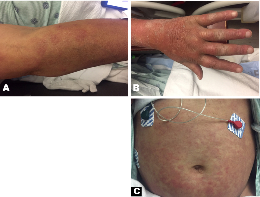
A 42-year-old Hispanic male presented to the hospital with a 2-year history of remitting and relapsing fever, diffuse maculopapular rash, and persistent generalized pain. Upon arrival, his vital signs were stable except for a fever of 39.3 C. He was alert and oriented, but complained of severe numbness and tingling in his hands and feet, chills, and night sweats accompanied by a diffuse, nonpruritic, painful maculopapular rash. Of note, he had been diagnosed with dermatomyositis four years ago given the constellation of findings. His medication history only consisted of prednisone 20 mg daily in order to manage his ‘dermatomyositis’. He reported similar episodes over the past two years and required an average of six hospitalizations per year to manage his symptoms. He characterized his pain as a burning sensation 8/10 in severity located across all of his joints, bones, and muscles that improved transiently with prednisone and morphine but was exacerbated by palpation and maintaining the same position for an extended period of time. His pain was accompanied by dyspnea and episodic vision loss 2–3 times per day that initially began as blurriness and progressed to excessive tear production and complete vision loss. These episodes would last 2-3 minutes and spontaneously resolve. On physical examination, the patient was in moderate distress and was found to have madarosis of bilateral eyebrows, sparse eyelashes, and ear helix with several non-tender flesh-colored and light brown papules (Figure 1). Numerous painful red indurated dermal papules and nodules were noted on his arms, forearms, lower abdomen, and upper back (Figure 2). Additionally, he had non-pitting edema in both hands, and his left foot had an overlying fourth metatarsal digit with a hyperkeratotic plantar plaque of approximately 1 x1.5 cm (Figure 3).
Due to his previous diagnosis of dermatomyositis, the patient was initially continued on his prednisone dose of 20 mg daily along with medication to manage pain. During his hospitalization, he had laboratory tests performed and was found to have leukocytosis, microcytic hypochromic anemia with iron level


DISCUSSION
Leprosy is a great mimicker of several rheumatologic diseases, and as a result, can be very challenging for primary physicians to diagnose [3]. This patient had been previously diagnosed with dermatomyositis. However, his diffuse maculopapular rash and pain did not respond to corticosteroids, whereas rheumatologic diseases typically respond to steroid therapy. The absence of gottron papules, shawl’s sign and heliotrope rash, negative antibodies and a normal CPK level decreases the likelihood of dermatomyositis in this setting. Establishing the true diagnosis of leprosy was complicated because the patient had atypical findings for leprosy, including painful maculopapular rash with no lepromas or hypoesthetic plaques. All findings were secondary to a rare presentation of leprosy known as ENL, which generally occurs in lepromatous leprosy cases and rarely in borderline cases. Young patients with high bacillary index and skin infiltration like the patient presented above are more prone to developing these reactions [5].
Although a vast majority of leprosy cases present with dermatologic and neurologic features, musculoskeletal manifestations are also quite common. Skin findings vary from macules to hypopigmented or hypoesthetic plaques and nodules, while neurologic manifestations can range from mononeuropathy to mononeuritis multiplex and distal symmetric polyneuropathy. The prevalence of rheumatic features in leprosy varies across case series, ranging from 1–2% described in large dermatology series to 60–80% in cases from rheumatology clinics. Charcot’s arthropathy is the classic rheumatologic manifestation of leprosy described in textbooks. It is usually found in longstanding cases with neurologic involvement; findings range from subluxation, dislocation, or pathologic fractures to complete joint destruction. In this case, the patient had an overlying fourth metatarsal digit [6],[7].
Leprosy may also mimic vasculitis, especially in cases with high bacillary load in the vascular endothelium. Commonly known as “lucio leprosy”, this variety is more common in Mexico and Costa Rica. Our patient had biopsy findings indicative of leukocytoclastic vasculitis, another factor contributing to difficulty differentiating from rheumatologic disease [8].
Leprosy is a great masquerader with manifestations resembling several different connective tissue diseases. Biopsy-proven cases of leprosy can present with heliotrope rash, muscle weakness, elevated muscle enzymes, oral ulcers, malar rash, and photosensitivity, but can also resemble scleroderma with Raynaud’s phenomenon, pittings scars, and skin thickening. In cases where findings don’t present with typical rheumatologic disease, skin biopsy should be performed to rule out destructive diseases such as leprosy and rare presentations such as ENL should be part of the differential. Demonstration of acid-fast bacilli in the dermis is the cornerstone of diagnosis [7],[8],[9].
CONCLUSION
Though leprosy poses a more significant disease burden in developing nations, it continues to have prevalence in developed regions. Diagnosis is often complicated by lack of awareness as well as atypical signs and symptoms. Therefore, it is critical that clinicians acknowledge these varied presentations and keep leprosy in their differential when faced with unusual findings reminiscent of rheumatologic disease.
REFERENCE
1.
Gupta L, Zanwar A, Wakhlu A, Agarwal V. Leprosy in the rheumatology clinic: An update on this great mimic. Int J Rheum Dis 2016;19(10):941–5.
[Pubmed]

2.
Andrade TCPC, Martins TY, Vieira BC, Santiago TM, Soares CT, Barreto JA. Lepromatous leprosy simulating rheumatoid arthritis - Report of a neglected disease. An Bras Dermatol 2017;92(3):389–91. [CrossRef]
[Pubmed]
3.
Kahawita IP, Walker SI, Lockwood DNJ. Leprosy type 1 reactions and erythema nodosum leprosum. An Bras Dermatol 2008;83(1):75–82.
4.
Costa PDSS, Fraga LR, Kowalski TW, Daxbacher ELR, Schuler-Faccini L, Vianna FSL. Erythema nodosum leprosum: Update and challenges on the treatment of a neglected condition. Acta Trop 2018;183:134–41. [CrossRef]
[Pubmed]
5.
Semwal S, Joshi D, Goel G, Mittal N, Majumdar K, Kapoor N. Cytological diagnosis of erythema nodosum leprosum in clinically unsuspected cases: A report of two cases. J Cytol 2018;35(1):63–5. [CrossRef]
[Pubmed]
6.
Haroon N, Agarwal V, Aggarwal A, Kumari N, Krishnani N, Misra R. Arthritis as presenting manifestation of pure neuritic leprosy: A rheumatologist's dilemma. Rheumatology (Oxford) 2007;46(4):653–6. [CrossRef]
[Pubmed]
7.
Chauhan S, Wakhlu A, Agarwal V. Arthritis in leprosy. Rheumatology (Oxford) 2010;49(12):2237–42. [CrossRef]
[Pubmed]
8.
Norman G, Joseph G. Richard J. Relapses in multibacillary patients treated with multi-drug therapy until smear negativity: Findings after twenty years. Int J Lepr Other Mycobact Dis 2004;72(1)1–7. [CrossRef]
[Pubmed]
9.
Rath D, Bhargava S, Kundu BK. Leprosy mimicking common rheumatologic entities: A trial for the clinician in the era of biologics. Case Rep Rheumatol 2014;2014:429698 [CrossRef]
[Pubmed]
SUPPORTING INFORMATION
Author Contributions
Marilyn Arosemena - Conception of the work, Design of the work, Revising the work critically for important intellectual content, Final approval of the version to be published, Agree to be accountable for all aspects of the work in ensuring that questions related to the accuracy or integrity of any part of the work are appropriately investigated and resolved.
Carlos I. Pacheco - Conception of the work, Design of the work, Revising the work critically for important intellectual content, Final approval of the version to be published, Agree to be accountable for all aspects of the work in ensuring that questions related to the accuracy or integrity of any part of the work are appropriately investigated and resolved.
Ankitha Lingamaneni - Acquisition of data, Drafting the work, Final approval of the version to be published, Agree to be accountable for all aspects of the work in ensuring that questions related to the accuracy or integrity of any part of the work are appropriately investigated and resolved.
Ankur Aneja - Acquisition of data, Drafting the work, Final approval of the version to be published, Agree to be accountable for all aspects of the work in ensuring that questions related to the accuracy or integrity of any part of the work are appropriately investigated and resolved.
Guarantor of SubmissionThe corresponding author is the guarantor of submission.
Source of SupportNone
Consent StatementOral informed consent was obtained from the patient for publication of this article.
Data AvailabilityAll relevant data are within the paper and its Supporting Information files.
Conflict of InterestAuthors declare no conflict of interest.
Copyright© 2019 Marilyn Arosemena et al. This article is distributed under the terms of Creative Commons Attribution License which permits unrestricted use, distribution and reproduction in any medium provided the original author(s) and original publisher are properly credited. Please see the copyright policy on the journal website for more information.


