|
Case Report
Unusual presentation of giant schwannoma in the sciatic nerve
1 Medical Sciences Course, Health Sciences School, Faculdade Ceres (FACERES), São José do Rio Preto, SP, Brazil
2 Neurosurgeon, Member of the Brazilian Society of Neurosurgery, Santa Mônica Hospital, Goiânia, GO, Brazil
3 Neurosurgeon, PhD in Neurosurgery, Member of the Brazilian Society of Neurosurgery, Professor at Santa Mônica Hospital, Goiânia, GO, Brazil
Address correspondence to:
Julia Brasileiro de Faria Cavalcante
Av. AnÃsio Haddad, 6751, Jardim Francisco Fernandes, São José do Rio Preto, SP,
Brazil
Message to Corresponding Author
Article ID: 100081Z06JC2020
Access full text article on other devices

Access PDF of article on other devices


How to cite this article
Cavalcante JBF, Cembraneli PN, Cavalcante RBF, Valente VF, Cavalcante JES. Unusual presentation of giant schwannoma in the sciatic nerve. Case Rep Int 2020;9:100081Z06JC2020.ABSTRACT
Introduction: Schwannomas are benign, encapsulated, slow-growing, generally solitary tumors originating from Schwann cells in the peripheral nerve heath. Schwannoma of the sciatic nerve is a rare condition and, therefore, little documented in the literature.
Case Report: A 77-year-old female patient arrived at the clinic complaining of pain and the appearance of a mass in the middle third of her left thigh. She presented with a well-defined mass, horizontally movable, hyperalgesia to the touch at the site of the mass, and positive Tinel’s sign. Magnetic resonance imaging showed multiple lesions suggestive of neurinomas, with intense nodular enhancement after gadolinium contrast injection, diffuse, in the right thigh and in the middle third of the left thigh. Surgical treatment was proposed by our team, but the patient preferred to continue the outpatient follow-up, because the injury did not interfere in her daily activities.
Conclusion: Although schwannoma in the sciatic nerve is rare, it should be considered in the differential diagnosis of patients presenting with conditions that affect this nerve.
Keywords: Schwannoma, Sciatic nerve, Tumor
INTRODUCTION
Peripheral nerve tumors are a heterogeneous group of benign tumors, which are rare in the general population [1]. Schwannomas are primary tumors of the neural sheath originating from Schwann cells [2],[3]. The occurrence of these tumors in the sciatic nerve is very rare (1%) [4]. These tumors grow slowly and since most of them are asymptomatic, they can be detected incidentally. It is also possible that they become symptomatic due to mechanical compression. Magnetic resonance imaging (MRI) is the diagnostic tool of choice for diagnosis of most soft-tissue tumors, including schwannomas, because of its high diagnostic predictability [5].
CASE REPORT
A 77-year-old female patient sought care due to persistent low-intensity pain and mass in the middle region of the left thigh for two years. She reported that the mass had been progressively growing in the last few months associated with paresthesia of the left lower limb.
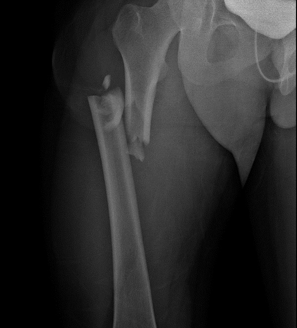
Physical examination showed preserved strength and hyperalgesia at the site of the mass. The patient described the pain as intermittent, radiating to the distal part of the left lower limb, with burning and electric shock-like sensations. Deep palpation of the left thigh region revealed a well-defined mass, approximately 8 cm in diameter (Figure 1), with regular contours, not hardened, horizontally movable, painful to the touch.
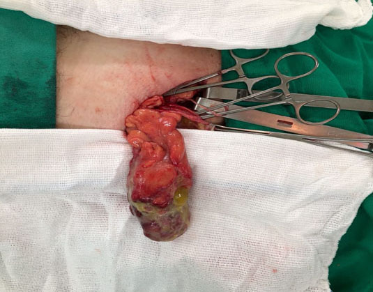
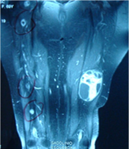
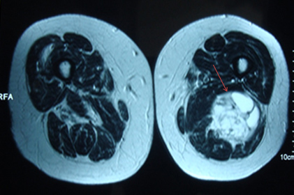
Magnetic resonance imaging showed multiple lesions suggestive of neurinomas, with intense nodular enhancement after gadolinium contrast injection, from 1.0 to 5.0 cm in diameter, diffuse, in the right thigh and in the middle of the left thigh, with heterogeneous hypersignal in T2 and foci of intralesional cystic degeneration (Figure 2). A tumor mass was visualized in the middle third of the sciatic nerve in the left thigh, measuring 9.0 × 7.0 × 6.0 cm, with cystic solid content (Figure 3).
The diagnosis of peripheral nerve schwannoma was established based on the characteristics of the MRI and the clinical history of the patient, who did not present other semiological findings. The surgical treatment was suggested by our team, but the patient refused it and chose to be followed up in the outpatient clinic, because she affirmed that the injury did not interfere in her daily activities.

DISCUSSION
Schwannomas of peripheral nerves in the lower limbs most frequently affect the posterior tibial nerve. The occurrence of this type of tumor in the sciatic nerve is very rare, with a frequency below 1%, and the presence of a painful palpable mass is the most reported clinical presentation [4],[6],[7].
Schwannomas grow very slowly over years. The lesions stretch slowly and lengthen the fascicles, still preserving the neurological function [8],[9]. Microscopically, the tumor is composed of spindled cells arranged in interlocking bundles. The nuclei tend to be arranged parallel to each other, showing an aspect that is called palisade arrangement [10].
Although most of these tumors are asymptomatic, they may be associated with pain and paresthesia in the corresponding neural territory. Hypoesthesia and motor deficits are more common in patients presenting with tumors larger than 4 cm in diameter [11],[12]. On physical examination, the tumor can be moved laterally, but not longitudinally toward the tumor trajectory, and percussion can produce positive Tinel’s sign [8],[9].
The exam of choice for the diagnosis and management of the lesion is MRI [5],[13], whose findings are: homogeneous mass, with medium to low signal intensity in T1-weighted sequences, while presenting hyperintense signal in T2 after gadolinium contrast infusion [9].
The treatment of schwannomas is surgical. The procedure aims at the complete resection of the tumor employing microsurgical techniques [8],[9],[13].
CONCLUSION
Although schwannoma in the sciatic nerve is a rare tumor, it should be considered in the differential diagnosis of patients presenting with conditions that affect this nerve.
REFERENCE
1.
Kang HJ, Shin SJ, Kang ES. Schwannomas of the upper extremity. J Hand Surg Br 2000;25(6):604–7. [CrossRef]
[Pubmed]

2.
Dwyer CL, Creager AJ, Lubahn JD. An unusual presentation of a digital schwannoma: Case report. Hand (N Y) 2015;10(2):301–4. [CrossRef]
[Pubmed]
3.
Kütahya H, Güleç A, Güzel Y, Kacira B, Toker S. Schwannoma of the median nerve at the wrist and palmar regions of the hand: A rare case report. Case Rep Orthop 2013;2013:950106. [CrossRef]
[Pubmed]
4.
Omezzine SJ, Zaara B, Ali MB, Abid F, Sassi N, Hamza HA. A rare cause of non discal sciatica: Schwannoma of the sciatic nerve. Orthop Traumatol Surg Res 2009;95(7):543–6. [CrossRef]
[Pubmed]
5.
Radojkovic M, Mihailovic D, Stojanovic M, Radojković D. Large retroperitoneal schwannoma: A rare cause of chronic back pain. J Int Med Res 2018;46(8):3404– 10. [CrossRef]
[Pubmed]
6.
Tan LA, Bradbury J, Bonnin J, Horn EM. Minimally invasive resection of an extrapelvic sciatic schwannoma. J Clin Neurosci 2010;17(10):1314–6. [CrossRef]
[Pubmed]
7.
Eroglu U, Bozkurt M, Ozates O, Akturk S, Tuna H. Sciatic nerve schwannoma: Case report. Turk Neurosurg 2014;24(1):120–2. [CrossRef]
[Pubmed]
8.
Friedman AH, Kline DG, Kim DH, Kitagawa RS. Benign tumors of the peripheral nerve. In: Winn RH, editor. Youmans Neurological Surgery. Volume 4. 6ed. Philadelphia, PA: Elsevier Saunders; 2011. p. 2518–21.
9.
Adani R, Tarallo L, Mugnai R, Colopi S. Schwannomas of the upper extremity: Analysis of 34 cases. Acta Neurochir (Wien) 2014;156(12):2325–30. [CrossRef]
[Pubmed]
10.
Bhattacharya JB, Singh M, Jain SL. Intraparotid schwannoma masquerading as primary spindle cell tumour of parotid: A diagnostic pitfall. J Cytol 2017;34(4):221–3. [CrossRef]
[Pubmed]
11.
Baderca F, Cojocaru S, Lazăr E, et al. Schwannoma of the lip: Case report and review of the literature. Rom J Morphol Embryol 2008;49(3):391–8.
[Pubmed]
12.
Hamdi MF, Aloui I, Ennouri Kh. Sciatica secondary to sciatic nerve schwannoma. Neurol India 2009;57(5):685–6. [CrossRef]
[Pubmed]
13.
Adani R, Baccarani A, Guidi E, Tarallo L. Schwannomas of the upper extremity: Diagnosis and treatment. Chir Organi Mov 2008;92(2):85–8. [CrossRef]
[Pubmed]
SUPPORTING INFORMATION
Author Contributions
Julia Brasileiro de Faria Cavalcante - Conception of the work, Design of the work, Acquisition of data, Analysis of data, Drafting the work, Revising the work critically for important intellectual content, Final approval of the version to be published, Agree to be accountable for all aspects of the work in ensuring that questions related to the accuracy or integrity of any part of the work are appropriately investigated and resolved.
Pedro Nogarotto Cembraneli - Conception of the work, Design of the work, Acquisition of data, Analysis of data, Drafting the work, Revising the work critically for important intellectual content, Final approval of the version to be published, Agree to be accountable for all aspects of the work in ensuring that questions related to the accuracy or integrity of any part of the work are appropriately investigated and resolved.
Renata Brasileiro de Faria Cavalcante - Conception of the work, Design of the work, Acquisition of data, Analysis of data, Drafting the work, Revising the work critically for important intellectual content, Final approval of the version to be published, Agree to be accountable for all aspects of the work in ensuring that questions related to the accuracy or integrity of any part of the work are appropriately investigated and resolved.
Volmer Fernandes Valente Junior - Conception of the work, Design of the work, Acquisition of data, Analysis of data, Drafting the work, Revising the work critically for important intellectual content, Final approval of the version to be published, Agree to be accountable for all aspects of the work in ensuring that questions related to the accuracy or integrity of any part of the work are appropriately investigated and resolved.
José Edison da Silva Cavalcante - Conception of the work, Design of the work, Acquisition of data, Analysis of data, Drafting the work, Revising the work critically for important intellectual content, Final approval of the version to be published, Agree to be accountable for all aspects of the work in ensuring that questions related to the accuracy or integrity of any part of the work are appropriately investigated and resolved.
Guarantor of SubmissionThe corresponding author is the guarantor of submission.
Source of SupportNone
Consent StatementWritten informed consent was obtained from the patient for publication of this article.
Data AvailabilityAll relevant data are within the paper and its Supporting Information files.
Conflict of InterestAuthors declare no conflict of interest.
Copyright© 2020 Julia Brasileiro de Faria Cavalcante et al. This article is distributed under the terms of Creative Commons Attribution License which permits unrestricted use, distribution and reproduction in any medium provided the original author(s) and original publisher are properly credited. Please see the copyright policy on the journal website for more information.