|
Case Report
Bouveret’s syndrome: An unusual cause of gastric outlet obstruction
1 General Surgery Department, Hospital Santa Luzia Elvas, Unidade Local Saúde Norte Alentejano, Portugal
2 General Surgeon, General Surgery Department, Hospital Santa Luzia Elvas, Unidade Local Saúde Norte Alentejano, Portugal
3 Gastroenterologist, Special Exams Unit (Gastroenterology), Hospital Santa Luzia Elvas, Unidade Local Saúde Norte Alentejano, Portugal
4 Radiologist, Radiology Department, Hospital Santa Luzia Elvas, Unidade Local Saúde Norte Alentejano, Portugal
5 Internal Medicine, Internal Medicine Department, Hospital Santa Luzia Elvas, Unidade Local Saúde Norte Alentejano, Portugal
6 Head of Internal Medicine, Internal Medicine Department, Hospital Santa Luzia Elvas, Unidade Local Saúde Norte Alentejano, Portugal
7 Head of Surgery Department, General Surgery Department, Hospital Santa Luzia Elvas, Unidade Local Saúde Norte Alentejano, Portugal
Address correspondence to:
Marta Reia
General Surgery Department, Hospital Santa Luzia, Elvas - Unidade Local Saúde do Norte Alentejano, Av. Mariana Martins, 7350-954 Elvas,
Portugal
Message to Corresponding Author
Article ID: 100099Z06MR2021
Access full text article on other devices

Access PDF of article on other devices


How to cite this article
Reia M, Messias F, Aguero C, Godinho R, Rodrigues T, Carvajal E, Cruz C, Urbano J, Silva V, Santos CP. Bouveret’s syndrome: An unusual cause of gastric outlet obstruction. Case Rep Int 2021;10:100099Z06MR2021.ABSTRACT
Introduction: Bouveret’s syndrome is a rare cause of pylorus or duodenal obstruction. This syndrome represents only 1–3% of all cases of gallstone ileus, more frequent in women and in the elderly. The clinical presentation is quite nonspecific, but in most cases the symptomatology suggests an upper digestive obstruction.
Case Report: We report a case of a 46-year-old man admitted with upper digestive obstruction due to a large biliary calculus that eroded through the gallbladder’s wall and pylorus, leading to digestive obstruction at this level. After attempted and unsuccessful endoscopic therapy, the patient was treated by surgery, with gastrotomy and gallstone removal. The fistula between the gallbladder and pylorus was not approached, because of the extent of the local inflammatory process, and elevated risk of complications.
Conclusion: Despite of being a benign disease, the morbidity and mortality of this syndrome may be elevated. The main objective of the treatment is to resolve the digestive obstruction, which can be achieved by upper gastrointestinal (GI) endoscopy, but most patients require a surgical approach. The treatment of the cholecystoduodenal/cholecystopyloric fistula—one-or two-stage treatment—is controversial, depending on the local inflammation, fibrosis, and anatomic distortion, as well the clinical condition of the patient.
Keywords: Bouveret’s syndrome, Duodenal obstruction, Gallstone ileus, Pylorus obstruction, Upper digestive obstruction
INTRODUCTION
Bouveret’s syndrome is a rare form of gallstone ileus, defined as a cholecysto-pyloric/duodenal or choledocho-pyloric/duodenal fistula with the passage of a big gallstone into the pylorus or duodenum, where the stone(s) obstruct gastroduodenal drainage [1],[2],[3],[4].
Fistula formation occurs due to pressure necrosis of the gallbladder wall and the adjacent organ (duodenum in this case) densely adhered to it. Other contributory factors include chronic inflammatory changes in the gallbladder wall and impaction of the stone in the gallbladder neck [5],[6],[7].
Due to the low specificity of symptoms and rarity of occurrence, the disease is easily misdiagnosed [3],[4] unless a high clinical suspicion is present.
The upper GI endoscopy and computed tomography (CT) of the abdomen are the tools that have proven useful for diagnosing this disease [1],[3],[8],[9].
This case report shows a patient who was successfully treated by surgery after unsuccessful endoscopic therapy.
CASE REPORT
A 46-year-old male patient, with medical history of hypertension, alcoholic, and smoking habits, who recurred to the emergency department with nausea and vomiting for the last 48 h. During the observation, the patient presented with upper GI hemorrhage, in the form of melena. The nasogastric drainage was abundant, with 4 L of dark hematic and stasis content.
On clinical examination, his vital signs were stable, there were no signs of hypovolemic shock or systemic inflammatory response, and his abdomen was soft, with no tenderness.
The blood tests showed a mild anemia, with hemoglobin of 11.4 g/dL (basal value of 15.4 g/dL), normocytic, normochromic, with a decreased hematocrit of 36.7%; the white blood cell count was 21.4 × 10⁹/L with neutrophilia (79.8%), total and direct bilirubin, alanine, and aspartate aminotransferase were normal, and the C-reactive protein was 6.49 mg/L, slightly elevated.
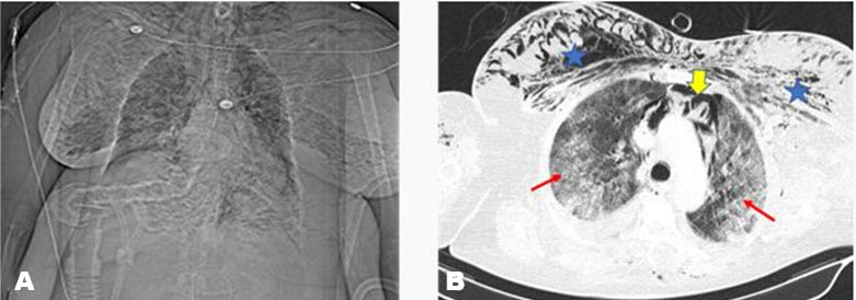
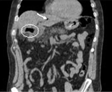
Abdominal radiography showed aerobilia and gastric dilation. The abdominal CT scan showed pneumobilia, with a choledocho-pyloric fistula, significant thickening of the duodenum wall, and large gallstone obstruction; there was no pneumoperitoneum or free intraperitoneal fluid (Figure 1 and Figure 2).
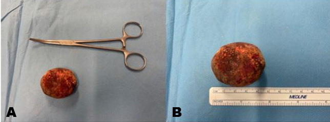
An upper GI endoscopy was performed, showing a large gallstone impacted in the pylorus, that could not be extracted endoscopically (Figure 3).
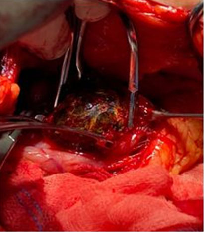
The patient was submitted to surgical treatment, laparotomy, followed by gastrotomy, gallstone retrieval, and gastrorrhaphy. The gallbladder was not approached due to exuberant inflammatory fibrosis and the elevated risk of biliary injury (Figure 4 and Figure 5).
The surgery occurred without complications, with minor blood loss. However, the post-operative blood tests showed a significant decrease in the hemoglobin level, of 7.7 g/dL. This downward evolution of the hemoglobin levels demonstrate the intensity of the digestive hemorrhage, regardless of other factors related to the surgery and fluid resuscitation that may impact on the hemogram.
There were no other post-operative complications, and the patient was discharged six days after surgery.
The definitive treatment (second-stage) was proposed to the patient, which consisted on cholecystectomy and fistula closure, but the patient decided to decline it, despite being informed about the related late possible complications. To the present day, the patient had no recurrent symptoms related to the cholecystopyloric fistula.


DISCUSSION
Bouveret’s syndrome is a rare cause of pylorus or duodenal obstruction. This syndrome represents only 1–3% of all cases of gallstone ileus, and is a distinct form of gallstone ileus [2],[5],[8],[10]. The typical gallstone ileus involves the small intestine (the most common location is the terminal portion of ileum (84%)), whereas in Bouveret’s syndrome the stomach (pylorus) or duodenum is involved [11].
Physiopathologically, this syndrome is characterized by the chronic inflammation of the gallbladder or biliary duct, with increase in intraluminal pressure followed by wall ischemia and subsequent perforation and fistula to the adjacent organ—communication between the biliary system and the GI tract—which allows the passage of gallstones from the gallbladder to the pylorus or duodenum [5],[6],[7].
While the majority of patients with gallstones do well, a small percentage of patients (approximately 6%) develop complications such as cholecysto-pylorus/duodenal or choledocho-pyloric/duodenal fistulas [11].
The risk factors for developing this syndrome include age greater than 70 years, female gender, previous cholecystitis, gallstones larger than 2–8 cm, and post-surgical altered GI anatomy [1],[3].
Bouveret’s syndrome was first reported in the literature by Beaussier in 1770, and was later named after French physician Leon Bouveret reported two cases in 1896 [5],[11].
Only 315 cases have been reported in the literature between 1967 and 2016 [8]. Bouveret’s syndrome tends to occur more commonly in women (65%) with a median age of 74.1 years at presentation [11]. As Bouveret’s syndrome often presents in patients with advanced age and multiple comorbidities, it is associated with a high morbidity and mortality rates (4.5–25%) [4],[9] [10].
The disease is easily misdiagnosed because of the low specificity of symptoms and rarity of occurrence. Symptoms like nausea and vomiting (86%), abdominal pain (71%), hematemesis (15%), melena, loss of weight, and anorexia have been described [3],[4]].
The classic specific Rigler’s triad is only present in 30–35% of the cases, and includes aerobilia, upper bowel obstruction, and an ectopic radiopaque stone [1],[2],[3],[5],[6],[8].
On physical exam, patients may present without abdominal pain, or may otherwise have abdominal tenderness, abdominal distention, and dehydration [7],[8],[12].
In our case, the patient’s vital signs were stable and the abdomen was soft, with no tenderness.
The blood test may show leukocytosis, deranged liver function tests, and elevation of inflammatory markers such as C-reactive protein [1],[7],[12]. In the present case, the patient had leukocytosis and a slightly increase C-reactive protein (6.49 mg/L).
The first diagnostic exam would be an abdominal radiograph, but it plays a limited role for diagnosing this disease since most of the gallstones are radiolucent [9], which may preclude the implementation of the Rigler’s triad, as it contemplates the presence of radiopaque gallstones. In our case, the abdominal radiography found aerobilia and gastric dilation.
The abdominal CT scan is more useful, diagnostic in about 60% of cases, with sensitivity of 93% and specificity of 100% [5],[9]. It is helpful in demonstrating the exact level of the obstruction, the biliary site of the duodenal fistula, and the status of the gallbladder [12]. However, approximately 15–25% of gallstones are isoattenuating and not well visualized on CT [11].
In our case, the abdominal CT scan showed pneumobilia with a choledocho-pyloric fistula, significant wall thickening of the duodenum, a large gallstone obstructing, and no sign of pneumoperitoneum of free intraperitoneal fluid.
The diagnosis of Bouveret’s syndrome is usually made via upper GI endoscopy, which is mandatory in this disease [6]. The first line of treatment should be endoscopic, with an attempt to retrieve the stone, despite the success rate of this procedure of 30–40%, approximately [9].
The visualization of the gallstone is only possible in about 69% of the cases, made more difficult in the presence of alimentary remaining or the non-progression of gastroscope beyond the pylorus [3].
In high-risk patients, in which surgical treatment comprises a greater risk, alternative non-invasive techniques were attempted, such as endoscopic lithotripsy, extracorporeal shock wave lithotripsy, electro-hydraulic lithotripsy, and endoscopic laser lithotripsy [11].
The endoscopic treatment and percutaneous lithotripsy should be attempted as the first approach in elderly patients, with multiple comorbidities. In general, the success rate of endoscopic extraction is dependent on stone size [8],[10].
Gallstones larger than 2.5 cm are more difficult to extract endoscopically, and have a greater risk ischemic ulceration of the adjacent pylorus or duodenal wall [11]. Despite being considered the initial treatment modality, some authors defend that endoscopic lithotomy increases the risk of esophageal injury, digestive tract perforation, and gastrointestinal bleeding [10].
When endoscopic treatment fails, surgical treatment is required. Currently, there are three surgical options described in the literature, without consensus on the indicated one. These procedures are lithotomy only, one-stage lithotomy plus cholecystectomy plus fistula closure, and two-stage lithotomy with differed cholecystectomy and fistula closure [13].
The criteria for the choice of the procedure are mainly clinic, regarding the patient’s age, comorbidities, clinical presentation of the syndrome, time of evolution and its effect on the patient clinical status. Consideration about the size of the calculi and fistula, its anatomic location and local inflammatory changes must also be taken in account [14].
In one-stage surgery, a combination of enterolithotomy (removal of the stone) plus cholecystectomy and fistula repair is performed. As advantage, this method prevents the need for a second operation and the occurrence of retrograde biliary tract infection, gallbladder cancer, and recurrence of gallstone ileus [15]. Described in literature, we find this approach applied in about 36% of the cases [3]. However, it is associated with significant morbidity—up to 66%—and mortality of 20–30%, and should only be attempted in stable and younger patients [3], [16], with better performance status and better physiologic reserve, or with a lesser degree of local inflammation or fibrosis [3]. The fact that the majority of patients with Bouveret’s syndrome are elderly, with a long history of systemic diseases, also contributes for a high incidence of postoperative complications and mortality [3],[7].
In the remaining 64% of the cases, a two-stage surgery strategy is chosen [3]. In the attempt to reduce the incidence of complications, in patients several times with high surgical risk and impaired physiologic status, the first stage of the surgical treatment comprehends a gastrotomy or duodenotomy with gallstone removal, without cholecystectomy or fistula repair. In case of Bouveret’s syndrome, if the local conditions allow it, the calculi should be mobilized toward the stomach, and removed through a gastrotomy; alternatively, they may be retrieved through a duodenal longitudinal incision, with emphasis on its closure to avoid stenosis [17]. The mortality of this staged approach is estimated to be around 12% [1],[3],[5],[7],[11],[12].
The definitive/second stage surgical treatment is differed until the clinical conditions of the patient are optimized, usually 2 to 3 months after the primary procedure, after the local inflammatory process is attenuated. The objectives in this stage are the removal of the gallbladder and any remaining calculus, and the cholecystoduodenal or cholecystopyloric fistula repair. An intraoperative cholangiogram may be useful for biliary anatomy clarification, and to assess the presence of residual biliary calculus on the biliary ducts, with possible lithotomy.
Alternatively, a simple enterolithotomy may be considered in frail patients, without any further intervention. In a meta-analysis of 1001 cases of gallstone ileus, only 10% of the patients required a second surgery for the repair of the biliary fistula, mandated by symptoms persistence [3],[18]. Despite simple lithotomy has long been associated with a lower mortality [17], this approach should not be generalized, because of the possible late complications that may ensue, and already discussed.
In the reported case, the upper endoscopy found a large gallstone impacted in the pylorus, which could not be extracted endoscopically, and surgical treatment was needed. A laparotomy was then performed, with gastrotomy, removal of the impacted stone, and gastrorrhaphy. The gallbladder was not approached as referred before [2],[5],[8],[11].
CONCLUSION
This case report illustrates a rare disease associated with high morbidity and mortality, which requires a high index of clinical suspicion for timely diagnosis. Bouveret’s syndrome represents 1–3% of all the cases of gallstone ileus, making it an exclusion diagnosis, after other more common causes are discarded. Treatment should be individualized according to the patient’s age, physical condition, and disease characteristics. The standard treatment is endoscopic. Despite prior case series have reported modest success rates with endoscopic treatment (30–40%), it is likely that endoscopic success rates are much higher now, as more innovative endoscopic techniques are used to treat these patients. In cases when the endoscopic treatment fails, surgery is indicated for the resolution of the upper gastrointestinal obstruction, accomplished by the gallstone removal.
REFERENCE
1.
Turner AR, Kudaravalli P, Ahmad H. Bouveret Syndrome. In: StatPearls. Treasure Island (FL): StatPearls Publishing; 2021.

2.
Castro S, Nadal A, Puerta S, et al. Síndrome de Bouveret, cartas al director. Cir Esp 2006;79(3):193–5. [CrossRef]
3.
Peixoto R, Correia, J, Soares MG, Gouveia A. Bouveret’s syndrome: A case report and a brief literature review. [Article in Portuguese]. Acta Med Port 2020;33(5):347–9. [CrossRef]
[Pubmed]
4.
Smith Z, Totten J, Hughes A, Strote J. Delayed diagnosis of gastric outlet obstruction from bouveret syndrome in a young woman. West J Emerg Med 2015;16(1):151–3. [CrossRef]
[Pubmed]
5.
Siadol Guerrero S, Canall Daza F, Jiménez Sánchez HC, Martínez Montalvo CM, Osorio Santos M. Case report of Bouveret syndrome: A strange cause of upper intestinal obstruction. Rev Colomb Gastroenterol 2019;34(4):445–9. [CrossRef]
6.
Ruiz-Santiago CM, Roldán-Ortiz S, Rodrigues-Olmo S, Soria-De La Cruz MJ, Reina-Cubero R. Síndrome de Bouveret: casos clínicos y revisión en la literatura. RAPD Online 2013;36(6).
7.
Mishra A, Jain A, Lal P, Hadke NS. Bouveret syndrome: A case report and review. J Gastroint Dig Sys 2013;3:133. [CrossRef]
8.
Wang F, Du ZQ, Chen YL, Chen TM, Wang Y, Zhou XR. Bouveret syndrome: A case report. World J Clin Cases 2019;7(23):4144–9. [CrossRef]
[Pubmed]
9.
Gajendran M, Muniraj T, Gelrud A. A challenging case of gastric outlet obstruction (Bouveret’s syndrome): A case report. J Med Case Rep 2011;5:497. [CrossRef]
[Pubmed]
10.
Caldwell K, Seeyuen L, Leggett P, et al. Bouveret syndrome: Current management strategies. Clin Exp Gastroenterol 2018;11:69–75. [CrossRef]
[Pubmed]
11.
Doycheva I, Limaye A, Suman A, Forsmark CE, Sultan S. Bouveret’s syndrome: Case report and review of the literature. Gastroenterol Res Pract 2009;2009:914951. [CrossRef]
[Pubmed]
12.
Nickel F, Müller-Eschner MM, Chu J, von Tengg-Kobligk H, Müller-Stich BP. Bouveret’s syndrome: Presentation of two cases with review of the literature and development of a surgical treatment strategy. BMC Surg 2013;13:33. [CrossRef]
[Pubmed]
13.
Nuño-Guzmán CM, Marín-Contreras ME, Figueroa-Sánchez M, Corona JL. Gallstone ileus, clinical presentation, diagnostic and treatment approach. World J Gastrointest Surg 2016;8(1):65–76. [CrossRef]
[Pubmed]
14.
Chica LFÁ, Cuéllar WB, Cardozo OLR. Biliary fistula and Bouveret’s syndrome. The same but different two case descriptions and a literature review. Rev Col Gastroenterol 2010;25(1):86–93.
15.
Lassandro F, Romano S, Ragozzino A, et al. Role of helical CT in diagnosis of gallstone ileus and related conditions. AJR Am J Roentgenol 2005;185(5):1159–65. [CrossRef]
[Pubmed]
16.
Rodríguez-Sanjuán JC, Casado F, Fernández MJ, Morales DJ, Naranjo A. Cholecystectomy and fistula closure versus enterolithotomy alone in gallstone ileus. Br J Surg 1997;84(5):634–7.
[Pubmed]
17.
Abou-Saif A, Al-Kawas FH. Complications of gallstone disease. Mirizzi syndrome, cholecystocholedochal fistula, and gallstone ileus. Am J Gastroenterol 2002;97(2):249–54. [CrossRef]
[Pubmed]
18.
Reisner RM, Cohen JR. Gallstone ileus: A review of 1001 reported cases. Am Surg 1994;60(6):441–6.
[Pubmed]
SUPPORTING INFORMATION
Author Contributions
Marta Reia - Conception of the work, Design of the work, Acquisition of data, Analysis of data, Drafting the work, Revising the work critically for important intellectual content, Final approval of the version to be published, Agree to be accountable for all aspects of the work in ensuring that questions related to the accuracy or integrity of any part of the work are appropriately investigated and resolved.
Francisco Messias - Conception of the work, Design of the work, Acquisition of data, Analysis of data, Drafting the work, Revising the work critically for important intellectual content, Final approval of the version to be published, Agree to be accountable for all aspects of the work in ensuring that questions related to the accuracy or integrity of any part of the work are appropriately investigated and resolved.
Coral Aguero - Conception of the work, Design of the work, Acquisition of data, Analysis of data, Drafting the work, Revising the work critically for important intellectual content, Final approval of the version to be published, Agree to be accountable for all aspects of the work in ensuring that questions related to the accuracy or integrity of any part of the work are appropriately investigated and resolved.
Rogério Godinho - Conception of the work, Design of the work, Acquisition of data, Analysis of data, Drafting the work, Revising the work critically for important intellectual content, Final approval of the version to be published, Agree to be accountable for all aspects of the work in ensuring that questions related to the accuracy or integrity of any part of the work are appropriately investigated and resolved.
Tiago Rodrigues - Conception of the work, Design of the work, Acquisition of data, Analysis of data, Drafting the work, Revising the work critically for important intellectual content, Final approval of the version to be published, Agree to be accountable for all aspects of the work in ensuring that questions related to the accuracy or integrity of any part of the work are appropriately investigated and resolved.
Eduardo Carvajal - Conception of the work, Design of the work, Acquisition of data, Analysis of data, Drafting the work, Revising the work critically for important intellectual content, Final approval of the version to be published, Agree to be accountable for all aspects of the work in ensuring that questions related to the accuracy or integrity of any part of the work are appropriately investigated and resolved.
Carlos Cruz - Conception of the work, Design of the work, Acquisition of data, Analysis of data, Drafting the work, Revising the work critically for important intellectual content, Final approval of the version to be published, Agree to be accountable for all aspects of the work in ensuring that questions related to the accuracy or integrity of any part of the work are appropriately investigated and resolved.
Juan Urbano - Conception of the work, Design of the work, Acquisition of data, Analysis of data, Drafting the work, Revising the work critically for important intellectual content, Final approval of the version to be published, Agree to be accountable for all aspects of the work in ensuring that questions related to the accuracy or integrity of any part of the work are appropriately investigated and resolved.
Victor Silva - Conception of the work, Design of the work, Acquisition of data, Analysis of data, Drafting the work, Revising the work critically for important intellectual content, Final approval of the version to be published, Agree to be accountable for all aspects of the work in ensuring that questions related to the accuracy or integrity of any part of the work are appropriately investigated and resolved.
Carlos Penalva Santos - Conception of the work, Design of the work, Acquisition of data, Analysis of data, Drafting the work, Revising the work critically for important intellectual content, Final approval of the version to be published, Agree to be accountable for all aspects of the work in ensuring that questions related to the accuracy or integrity of any part of the work are appropriately investigated and resolved.
Guarantor of SubmissionThe corresponding author is the guarantor of submission.
Source of SupportNone
Consent StatementWritten informed consent was obtained from the patient for publication of this article.
Data AvailabilityAll relevant data are within the paper and its Supporting Information files.
Conflict of InterestAuthors declare no conflict of interest.
Copyright© 2021 Marta Reia et al. This article is distributed under the terms of Creative Commons Attribution License which permits unrestricted use, distribution and reproduction in any medium provided the original author(s) and original publisher are properly credited. Please see the copyright policy on the journal website for more information.