|
Clinical Image
Electrocardiogram changes mimicking ventricular tachycardia, ventricular fibrillation and torsades de pointes in a patient with Parkinsonism
1 Professor of Cardiology, Akdeniz Sifa Hospital, Antalya, Turkey
Address correspondence to:
Mehmet Ozaydin
Akdeniz Sifa Konyaalti Medical Center, Kuskavagi Mah., Ataturk Blv. No: 81, 07070 Konyaalti, Antalya,
Turkey
Message to Corresponding Author
Article ID: 100100Z06MO2021
Access full text article on other devices

Access PDF of article on other devices


How to cite this article
Ozaydin M. Electrocardiogram changes mimicking ventricular tachycardia, ventricular fibrillation and torsades de pointes in a patient with Parkinsonism. Case Rep Int 2021;10:100100Z06MO2021.ABSTRACT
No Abstract
Keywords: Artifacts, Electrocardiogram changes, Parkinsonism, Ventricular arrhythmias
CASE REPORT
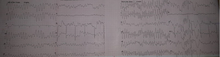
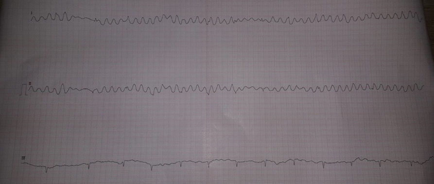
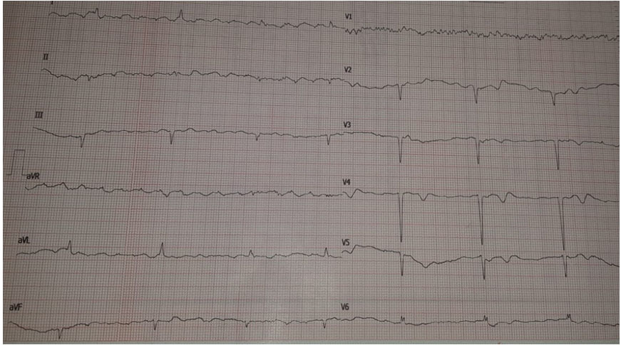
A 79-year-old man with hypertension, permanent atrial fibrillation, and status post-bypass surgery presented to emergency department with fatigue. The diagnosis of ventricular tachycardia (Figure 1) was made by the emergency physician. The patient was seen in emergency department as per planned cardiology consultation. On questioning it appeared that the patient had no palpitations but had the diagnosis of Parkinsonism. His blood pressure was 130/70 mmHg and pulse rate was 70 bpm with irregularly irregular rhythm. When electrocardiogram (ECG) was analyzed in detail, it was clear that ECG changes were secondary to tremor due to Parkinsonism, not ventricular tachycardia. He was hospitalized for rhythm follow-up. During hospitalization, further rhythm records showed that he had further artifacts mimicking torsades de pointes (Figure 2) and ventricular fibrillation (Figure 3). Electrocardiogram strips during minimal tremor showed that he has no ventricular arrhythmia but only atrial fibrillation (Figure 4). Tremor was more prominent in the right hand and all the recordings mimicking ventricular arrhythmias were best seen in all the extremity leads except DIII. On the other hand, tremor was less prominent in left hand and the artifacts were not seen in lead DIII (Figure 1 and Figure 3), which connects left hand to foot. All of these features indicated that his ECG changes were due to tremor-induced artifacts, but not ventricular arrhythmias.
If artifact is suspected to be responsible for ECG changes, the ECG should be re-taken after the underlying cause of artifact is corrected. The cause of artifact was tremor in this patient and repeated ECG during minimal tremor showed no ventricular arrhythmia-like appearance.

DISCUSSION
Artifacts may cause some ECG changes that look like rhythm disturbances. Tremor, electromagnetic interference and cell phones are the most common causes of artifacts. Several previous reports have indicated that tremor may cause ECG or telemetry changes mimicking arrhythmia [1],[2],[3],[4]. Malik and Nahed [1] presented telemetry changes mimicking polymorphic ventricular tachycardia in a patient with coronary artery disease and Parkinson’s disease undergoing glioma resection. Gunawardena [2] and Bergin and Lynch [3] reported ECG changes mimicking torsades de pointes. In another case report, atrial flutter/fibrillation or ventricular tachycardia like appearances were reported in a patient with tremor [4]. Here, we report a case of Parkinsonism presenting with tremor-induced ECG changes mimicking different types of ventricular arrhythmias.
CONCLUSION
Instead of just looking at laboratory test results, doctors have to talk to the patients about their symptoms and medical histories and also examine them in order to give the correct diagnosis because we need to treat the patients but not their laboratory findings.
REFERENCE
1.
Malik AN, Nahed BV. Tremor mimicking ventricular tachycardia on telemetry. World Neurosurg 2021;147:158–9. [CrossRef]
[Pubmed]

2.
Gunawardena I. Parkinson’s tremor mimicking torsades de pointes. Br J Hosp Med (Lond) 2011;72(10):593. [CrossRef]
[Pubmed]
3.
Bergin U, Lynch RM. Artefact mimicking torsades: Treat the patient not the ECG. Practitioner 2017;261(1801):23–6.
[Pubmed]
4.
Hwang WJ, Chen JY, Sung PS, Lee JC. Parkinsonian tremor-induced electrocardiographic artifacts mimicking atrial flutter/fibrillation or ventricular tachycardia. Int J Cardiol 2014;173(3):597–600. [CrossRef]
[Pubmed]
SUPPORTING INFORMATION
Author Contributions
Mehmet Ozaydin - Conception of the work, Design of the work, Acquisition of data, Analysis of data, Drafting the work, Revising the work critically for important intellectual content, Final approval of the version to be published, Agree to be accountable for all aspects of the work in ensuring that questions related to the accuracy or integrity of any part of the work are appropriately investigated and resolved.
Guarantor of SubmissionThe corresponding author is the guarantor of submission.
Source of SupportNone
Consent StatementWritten informed consent was obtained from the patient for publication of this article.
Data AvailabilityAll relevant data are within the paper and its Supporting Information files.
Conflict of InterestAuthor declares no conflict of interest.
Copyright© 2021 Mehmet Ozaydin. This article is distributed under the terms of Creative Commons Attribution License which permits unrestricted use, distribution and reproduction in any medium provided the original author(s) and original publisher are properly credited. Please see the copyright policy on the journal website for more information.