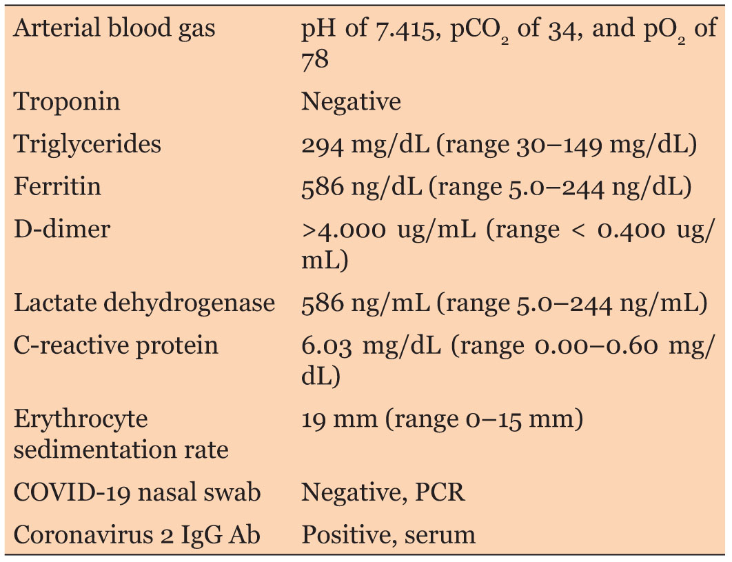
|
Clinical Image
The unilateral persistent nephrogram on CT IVP: Take your time
1 Resident, Department of Urology, Ikazia Hospital, Rotterdam, The Netherlands
2 Urologist, Department of Urology, Ikazia Hospital, Rotterdam, The Netherlands
3 Radiologist, Department of Radiology, Ikazia Hospital, Rotterdam, The Netherlands
4 Internist, Department of Internal Medicine, Ikazia Hospital, Rotterdam, The Netherlands
Address correspondence to:
Frederika Straten Straten
Gasthuissteeg 23B, Delft,
The Netherlands
Message to Corresponding Author
Article ID: 100103Z06FS2021
Access full text article on other devices

Access PDF of article on other devices


How to cite this article
Straten FJ, Nienhuis JE, de Jonge LCW, Dees A. The unilateral persistent nephrogram on CT IVP: Take your time. Case Rep Int 2021;10:100103Z06FS2021.ABSTRACT
No Abstract
Keywords: CT IVP, Persistent nephrogram, Renal colic
CASE REPORT
A 69-year-old Caucasian male patient consulted the Urology Department of our hospital because of macroscopic hematuria and left lower abdominal pain. There was no history of nephrolithiasis. Physical examination of the abdomen was normal, and ultrasound of the kidneys by the urologist showed no hydronephrosis. Cystoscopy was normal and during cystoscopy urine was clear. Urine cytology showed no signs of a highly grade urothelial cell neoplasia, urine culture was sterile, and urine microscopy showed more than 50 red blood cells per high power fields. To further analyze the hematuria, a computed tomography (CT) intravenous pyelogram (IVP), also called a CT intravenous urography (IVU), with furosemide administration was planned. A CT IVP was performed at 9 AM, and on that specific day the patient presented with renal colic. The CT scan showed normal contrast excretion of the right kidney and a persistent nephrogram on the left side, with no contrast within the pyelum and ureter (Figure 1). There were no signs of renal artery stenosis, renal vein thromboses or urolithiasis. At 4 PM, the CT scan was repeated, a slightly dilatated left pyelum and ureter filled with contrast material was seen, and there was some perirenal fat infiltration (Figure 2). There was a urinary tract obstruction causing the dilatation and the persistent nephrogram, and our differential diagnosis included a passed stone or pyelonephritis with thick pus in the ureter. The patient was admitted to the hospital for pain relief and was discharged home in good clinical condition the next day. Three months later, the CT IVP was repeated and a normal nephrogram was seen on both sides (Figure 3).


DISCUSSION
The nephrogram is the radiographic image of the contrast-filled renal parenchyma [1]. Normally three different phases of renal contrast enhancement follow each other after the administration of intravenous contrast. During the first phase, the corticomedullary phase, contrast material reaches the renal cortical capillaries resulting in a density differentiation between the cortex and medulla. Afterwards, the contrast material is filtered by the glomeruli and enters the renal tubules resulting in a homogenous appearance of the kidney, the so-called nephrographic phase. During the last phase, the excretory phase, the contrast is excreted into the calices causing the renal collecting system to appear [2]. Diuretic administration, for instance with furosemide, will enhance the glomerular filtration and the wash out of contrast. To produce a normal nephrogram adequate renal perfusion, normal glomerular filtration and normal tubular transit and function are required as well as the absence of a urinary tract obstruction [1],[3]. A persistent nephrogram, for instance 6 hours after the initial investigation, is an abnormal nephrogram defined as the renal retention of contrast material and may occur bilaterally or unilaterally [4]. Unilateral causes of a persistent nephrogram described in the literature include etiologies such as renal artery stenosis, renal vein thrombosis and urinary tract obstructions [1],[5],[6],[7]. Renal artery stenosis causes a unilateral decrease in the renal blood flow resulting in a decline in the glomerular filtration and tubular transit causing a prolongation of the nephrographic phase. In renal vein thromboses, the glomerular filtration and tubular transit are decreased due to the opposition of the arterial perfusion gradient as a result of the intravenous clot. Additionally, the development of edema will result in an increase in the parenchymal pressure causing a compression of the tubular lumina which further decreases the tubular transit. Obstruction of the urinary outflow increases the internal pressure proximal to the blockage which is transmitted in a retrograde direction to the level of the nephron, which opposes the forward perfusion gradient and therefore causes a decrease in the glomerular filtration and tubular transit. A urinary tract obstruction can be caused by a variety of urinary and extra urinary abnormalities (such as the presence of stones, blood clots, pus, and tumors, and with an extrinsic compression of the ureter), and usually presents with acute renal colic [7]. As to our patient: we assume that a small stone was responsible for his transient complaints and the obstructive findings.
CONCLUSION
Causes of a unilateral persistent nephrogram are a renal artery stenosis, a renal vein thrombosis and a urinary tract obstruction. Urinary tract obstruction is associated with renal colic and can be caused by a variety of urinary and extraurinary abnormalities. It is important to repeat the radiography after 6 hours to visualize the cause of urinary tract obstruction, especially in patients presenting with hematuria and colic.
REFERENCE
1.
Saunders HS, Dyer RB, Shifrin RY, Scharling ES, Bechtold RE, Zagoria RJ. The CT nephrogram: Implications for evaluation of urinary tract disease. Radiographics 1995;15(5):1069–85. [CrossRef]
[Pubmed]

2.
Yuh BI, Cohan RH. Different phases of renal enhancement: Role in detecting and characterizing renal masses during helical CT. AJR Am J Roentgenol 1999;173(3):747–55. [CrossRef]
[Pubmed]
3.
Dyer R, Munitz HA, Bechtold R, Choplin RH. The abnormal nephrogram. Radiographics 1986;6(6):1039–63. [CrossRef]
[Pubmed]
4.
Gagne SM, Newbury A, Nowitzki KM, Chen BY, Lo HS. Name that nephrogram: Asymmetric renal enhancement in the acute care setting. Curr Probl Diagn Radiol 2019;48(6):616–25. [CrossRef]
[Pubmed]
5.
Ibrahim MI, Qayyum A, Khan A, et al. Retroaortic left renal vein as a cause of unilateral dense persistent nephrogram. Pak Armed Forces Med J 2015;65(6):849–50.
6.
Mahajan A, Travis S. Case of the month# 170: The unilateral persistent nephrogram after endovascular aortic aneurysm repair (EVAR): New life in an old sign. Can Assoc Radiol J 2011;62(3):229–30. [CrossRef]
[Pubmed]
7.
Wolin EA, Hartman DS, Olson JR. Nephrographic and pyelographic analysis of CT urography: Principles, patterns, and pathophysiology. AJR Am J Roentgenol 2013;200(6):1210–4. [CrossRef]
[Pubmed]
SUPPORTING INFORMATION
Author Contributions
Frederika Straten Straten - Conception of the work, Design of the work, Acquisition of data, Analysis of data, Drafting the work, Revising the work critically for important intellectual content, Final approval of the version to be published, Agree to be accountable for all aspects of the work in ensuring that questions related to the accuracy or integrity of any part of the work are appropriately investigated and resolved.
Jaap E Nienhuis - Conception of the work, Design of the work, Acquisition of data, Analysis of data, Drafting the work, Revising the work critically for important intellectual content, Final approval of the version to be published, Agree to be accountable for all aspects of the work in ensuring that questions related to the accuracy or integrity of any part of the work are appropriately investigated and resolved.
Leenoud CW de Jonge - Acquisition of data, Analysis of data, Revising the work critically for important intellectual content, Final approval of the version to be published, Agree to be accountable for all aspects of the work in ensuring that questions related to the accuracy or integrity of any part of the work are appropriately investigated and resolved.
Adriaan Dees - Conception of the work, Design of the work, Acquisition of data, Analysis of data, Drafting the work, Revising the work critically for important intellectual content, Final approval of the version to be published, Agree to be accountable for all aspects of the work in ensuring that questions related to the accuracy or integrity of any part of the work are appropriately investigated and resolved.
Guarantor of SubmissionThe corresponding author is the guarantor of submission.
Source of SupportNone
Consent StatementWritten informed consent was obtained from the patient for publication of this article.
Data AvailabilityAll relevant data are within the paper and its Supporting Information files.
Conflict of InterestAuthors declare no conflict of interest.
Copyright© 2021 Frederika J Straten et al. This article is distributed under the terms of Creative Commons Attribution License which permits unrestricted use, distribution and reproduction in any medium provided the original author(s) and original publisher are properly credited. Please see the copyright policy on the journal website for more information.