|
Case Report
Atypical case of pulmonary Langerhans cell histiocytosis presenting as miliary nodules
1 Internal Medicine, MercyONE North Iowa, Mason City, IA, USA
2 Pulmonology, MercyONE North Iowa, Mason City, IA, USA
Address correspondence to:
Michelle Low
652 S Tennessee Pl, Mason City, IA 50401,
USA
Message to Corresponding Author
Article ID: 100106Z06ML2022
Access full text article on other devices

Access PDF of article on other devices


How to cite this article
Low M, Lasley K, Singh P. Atypical case of pulmonary Langerhans cell histiocytosis presenting as miliary nodules. Case Rep Int 2022;11:100106Z06ML2022.ABSTRACT
Pulmonary Langerhans cell histiocytosis (PLCH) is a rare smoking-related interstitial lung disease affecting primarily young adult smokers. Diagnosis is made with typical radiographic findings of centrilobular nodules and cystic lesions on high resolution computed tomography (HRCT) confirmed with tissue biopsy. We describe a case of a 59-year-old man presenting with chronic tobacco use and cough who was found to have an atypical radiographic finding of miliary nodules on HRCT that was confirmed as PLCH on transbronchial lung biopsy.
Keywords: Interstitial lung disease, Miliary nodules, Pulmonary Langerhans cell histiocytosis, Smoking
INTRODUCTION
Pulmonary Langerhans cell histiocytosis (PLCH) is a rare interstitial lung disease of clonal dendritic cell infiltration that primarily affects young adult smokers with peak incidence at 20–40 years of age [1]. Among biopsy-confirmed interstitial lung disease, PLCH comprises 4–5% of cases, and of those >90% have a history of tobacco use [2]. Alterations in mitogen-activated protein kinase (MAPK) including BRAF deletions have been observed in majority of lesions, and in refractory disease, identifying these aberrations can help guide targeted treatment [3]. The etiology has yet to be fully understood but is thought to be initiated by an inflammatory response to cigarette smoke with nodular and eventual cystic lung destruction [4]. As such, the radiographic findings of PLCH typically include granulomatous nodules and cystic lesions in the upper and middle lobes [5]. Symptoms range from asymptomatic to respiratory failure, but most commonly include nonproductive cough, dyspnea, fatigue, weight loss, and chest pain [5]. Definitive diagnosis is established with immunohistochemistry for >5% CD1a or CD207 positive cells obtained through bronchoalveolar lavage, lung biopsy, or cystic bone lesion biopsy [1],[6]. Herein we describe an atypical presentation of bilateral miliary nodules requiring transbronchial lung biopsy to diagnose PLCH.
CASE REPORT
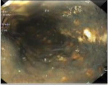
A 59-year-old nicotine-dependent man with reported history of adolescent asthma and chronic obstructive pulmonary disease was referred to pulmonology clinic following a chest X-ray with incidental new findings of upper lobe interstitial nodularity suggestive of granulomatous disease. Upon presentation to pulmonology clinic his only complaints were chronic cough for 10 years duration and exertional dyspnea. Given his radiographic findings high resolution computed tomographic (HRCT) examination and vasculitis workup was ordered. Blood work was only significant for elevated erythrocyte sedimentation rate (ESR) 80 mm/h, elevated C-reactive protein (CRP) 2.3 mg/dL, and elevated eosinophils 5.3% with 0.6×10³/uL absolute eosinophils. Tuberculosis Quantiferon test, vasculitis workup, and angiotensin converting enzyme (ACE) levels were within normal limits. High resolution computed tomography examination revealed innumerable tiny bilateral pulmonary nodules consistent with miliary pattern (Figure 1). Bronchoscopy with transbronchial lung biopsy was subsequently performed and found to contain focal areas of histiocytes associated with CD1a positive cells on immunohistochemistry, confirming PLCH (Figure 2). The patient was educated on the disease progression and thus strongly advised with smoking cessation. Exertional dyspnea symptoms were controlled with long-acting bronchodilator and inhaled corticosteroid.


DISCUSSION
Pulmonary Langerhans cell histiocytosis has been classically characterized by lesions that are bilateral, symmetric, and have upper lobe predominant nodules and cysts sparing the lung bases. Though the concurrence of centrilobular nodules and cysts sparing the lower lung fields is a diagnostic feature of PLCH, radiographic findings can vary depending on the stage of disease [7],[8]. Early stages of PLCH are characterized by bronchiolocentric nodules generally 1–10 mm in size and can be cavitating. As the disease progresses cysts predominate and can transform from thick-walled (>2 mm) to thin-walled (<2 mm), eventually resulting in endstage fibrocystic patterns remaining in the upper- and middle-lung fields [4],[8]. This spectrum of nodular and cystic changes along the course of disease makes PLCH difficult to diagnose on radiographic findings alone as it can mimic other pathologies including hematogenous spread of metastatic disease, primary malignancy, and miliary tuberculosis [6],[8].
This rare case of PLCH presenting as diffuse miliary nodules without co-existing cystic lesions demonstrates the importance of considering uncommon diagnoses in the face of unique lesions characteristic of other disease processes [8]. As evidenced in this case, it can be challenging to diagnose PLCH with non-characteristic lesions without a high index of suspicion, as it was only following extensive workup ruling out other etiologies that PLCH was confirmed on transbronchial lung biopsy.
Smoking cessation remains the most critical component of PLCH management but can include immunosuppressive and chemotherapies, though such treatment is not proven to reduce long-term mortality or improve lung function [9],[10]. Extrapulmonary manifestations of LCH include skeletal involvement as bone lytic lesions, dermatologic lesions, and central nervous system involvement most commonly diabetes insipidus. Overall prognosis is favorable as early smoking cessation can result in regression of the interstitial changes in early manifestations of PLCH [11]. However, complications can include initial manifestations of pneumothorax in approximately 15% of patients to pulmonary hypertension in >75% of symptomatic patients in advanced stages [2],[10],[12],[13],[14]. Publishing reports suggest a median survival of 10–12 years following the disease diagnosis.
CONCLUSION
Diagnosis of PLCH includes lung biopsy and radiographic findings on HRCT of centrilobular nodules and cystic lesions in various stages typically in the upper and middle lobes. Practicing physicians should be aware that a miliary distribution can be an atypical radiographic presentation of PLCH. PLCH as a result should be part of the differential diagnosis in a patient who tests negative for tuberculosis with miliary radiographic findings, and further investigation, including a biopsy of the lesion, should be pursued.
REFERENCE
1.
Tazi A. Adult pulmonary Langerhans’ cell histiocytosis. Eur Respir J 2006;27(6):1272–85. [CrossRef]
[Pubmed]

2.
Vassallo R, Ryu JH, Colby TV, Hartman T, Limper AH. Pulmonary Langerhans’-cell histiocytosis. N Engl J Med 2000;342(26):1969–78. [CrossRef]
[Pubmed]
3.
Jouenne F, Chevret S, Bugnet E, et al. Genetic landscape of adult Langerhans cell histiocytosis with lung involvement. Eur Respir J 2020;55(2):1901190. [CrossRef]
[Pubmed]
4.
Suri HS, Yi ES, Nowakowski GS, Vassallo R. Pulmonary Langerhans cell histiocytosis. Orphanet J Rare Dis 2012;7:16. [CrossRef]
[Pubmed]
5.
Kambouchner M, Basset F, Marchal J, Uhl JF, Hance AJ, Soler P. Three-dimensional characterization of pathologic lesions in pulmonary langerhans cell histiocytosis. Am J Respir Crit Care Med 2002;166(11):1483–90. [CrossRef]
[Pubmed]
6.
Sharma P, Dhingra KK, Sural S, Mandal AK, Singh T. Langerhans cell histiocytosis masquerading as tuberculosis: A diagnostic dilemma resulting in inappropriate anti-tubercular therapy. Pediatr Blood Cancer 2009;53(1):111–3. [CrossRef]
[Pubmed]
7.
Seely JM, Salahudeen Sr S, Cadaval-Goncalves AT, et al. Pulmonary Langerhans cell histiocytosis: A comparative study of computed tomography in children and adults. J Thorac Imaging 2012;27(1):65–70. [CrossRef]
[Pubmed]
8.
Castoldi MC, Verrioli A, De Juli E, Vanzulli A. Pulmonary Langerhans cell histiocytosis: The many faces of presentation at initial CT scan. Insights Imaging 2014;5(4):483–92. [CrossRef]
[Pubmed]
9.
Vassallo R, Ryu JH, Schroeder DR, Decker PA, Limper AH. Clinical outcomes of pulmonary Langerhans’-cell histiocytosis in adults. N Engl J Med 2002;346(7):484–90. [CrossRef]
[Pubmed]
10.
Radzikowska E. Update on pulmonary Langerhans cell histiocytosis. Front Med (Lausanne) 2021;7:582581. [CrossRef]
[Pubmed]
11.
Hagmeyer L, Randerath W. Smoking-related interstitial lung disease. Dtsch Arztebl Int 2015;112(4):43–50. [CrossRef]
[Pubmed]
12.
Mendez JL, Nadrous HF, Vassallo R, Decker PA, Ryu JH. Pneumothorax in pulmonary Langerhans cell histiocytosis. Chest 2004;125(3):1028–32. [CrossRef]
[Pubmed]
13.
Fartoukh M, Humbert M, Capron F, et al. Severe pulmonary hypertension in histiocytosis X. Am J Respir Crit Care Med 2000;161(1):216–23. [CrossRef]
[Pubmed]
14.
Chaowalit N, Pellikka PA, Decker PA, et al. Echocardiographic and clinical characteristics of pulmonary hypertension complicating pulmonary Langerhans cell histiocytosis. Mayo Clin Proc 2004;79(10):1269–75. [CrossRef]
[Pubmed]
SUPPORTING INFORMATION
Author Contributions
Michelle Low - Conception of the work, Design of the work, Acquisition of data, Analysis of data, Drafting the work, Revising the work critically for important intellectual content, Final approval of the version to be published, Agree to be accountable for all aspects of the work in ensuring that questions related to the accuracy or integrity of any part of the work are appropriately investigated and resolved.
Katharine Lasley - Conception of the work, Design of the work, Drafting the work, Final approval of the version to be published, Agree to be accountable for all aspects of the work in ensuring that questions related to the accuracy or integrity of any part of the work are appropriately investigated and resolved.
Pranav Singh - Conception of the work, Design of the work, Revising the work critically for important intellectual content, Final approval of the version to be published, Agree to be accountable for all aspects of the work in ensuring that questions related to the accuracy or integrity of any part of the work are appropriately investigated and resolved.
Guarantor of SubmissionThe corresponding author is the guarantor of submission.
Source of SupportNone
Consent StatementWritten informed consent was obtained from the patient for publication of this article.
Data AvailabilityAll relevant data are within the paper and its Supporting Information files.
Conflict of InterestAuthors declare no conflict of interest.
Copyright© 2022 Michelle Low et al. This article is distributed under the terms of Creative Commons Attribution License which permits unrestricted use, distribution and reproduction in any medium provided the original author(s) and original publisher are properly credited. Please see the copyright policy on the journal website for more information.