|
Case Report
Two cases of urinary schistosomiasis with unusual egg presentations: Dra 1 repeat sequence not detected
1 Lecturer II, Department of Microbiology, Faculty of Life Sciences, Ahmadu Bello University, Zaria, Kaduna State, Nigeria
2 Professor, Department of Microbiology, Faculty of Life Sciences, Ahmadu Bello University, Zaria, Kaduna State, Nigeria
3 Professor, Department of Public Health and Preventive Medicine, Faculty of Veterinary Medicine, Ahmadu Bello, University, Zaria, Kaduna State, Nigeria
Address correspondence to:
Henry Gabriel Bishop
Department of Microbiology, Ahmadu Bello University, Zaria, Kaduna State,
Nigeria
Message to Corresponding Author
Article ID: 100120Z06HB2023
Access full text article on other devices

Access PDF of article on other devices


How to cite this article
Bishop HG, Inabo HI, Ella EE, Bello M. Two cases of urinary schistosomiasis with unusual egg presentations: Dra 1 repeat sequence not detected. Case Rep Int 2023;12(2):10–14.ABSTRACT
Introduction: Schistosoma haematobium is the primary cause of urinary schistosomiasis in man. It is rare to find other human schistosome species in urine because they are located in the intestines, or those of animal origin. Mixed infections of human and animal species of schistosomes may occur in cattle breeding areas like Nigeria.
Case Report: During a prevalence study on urinary schistosomiasis, two teenage boys from different local government areas (LGAs) of Kaduna State, Nigeria had mixed urinary Schistosoma infections. Their urine samples were centrifuged at 3000 rpm (revolutions per minute) for 5 minutes. Microscopic examination of the urine sediments revealed highly polymorphic eggs (or morphotypes). After subjecting the genomic DNA for detection of S. haematobium Dra 1 tandem repeat sequence by polymerase chain reaction (PCR), it was not amplified. However, there was amplification in a classical urinary schistosomiasis caused by S. haematobium (which served as positive control).
Conclusion: Unusual egg presentations in urinary schistosomiasis may present a dilemma in making diagnostic conclusion. Hence, these two cases suggest the possibility of human–animal Schistosoma hybrids circulating in the area, especially S. haematobium–S. bovis hybrids.
Keywords: Kaduna State, Nigeria, Schistosoma bovis, Schistosoma haematobium
INTRODUCTION
Schistosomiasis is an acute and chronic parasitic disease caused by blood flukes of the genus Schistosoma [1]. The disease is most prevalent among people in rural and some urban areas of tropical and sub-tropical countries, especially those communities that lack access to safe drinking water and good sanitation [1],[2],[3]. Schistosomiasis is regarded as one of the neglected tropical diseases affecting man [4]. In Africa, the distribution of S. haematobium is sympatric with species infecting domestic cattle and wild ungulates, including S. bovis, S. intercalatum and other species that are closely related [5],[6]. Members of S. haematobium group are known to develop in the same Bulinus snails (as common intermediate hosts), producing almost indistinguishable cercariae [5],[7].
Children are mostly unaware of the risk of transmission of schistosomiasis via cercariae-infested water bodies and hence more infections occur [2]. There had been reports of hybrid schistosomes in some West African countries like Mali, Senegal, Benin, and Cameroon. These countries together with Nigeria share the River Niger and its tributaries. In Nigeria, control of schistosomiasis had been focused on preventing human contact with cercarial-infested water bodies, but there is lesser attention on the possible role of livestock as reservoir hosts [8]. In cattle-raising areas, as humans undertake agricultural, recreational, or fishing activities in the water bodies, cattle also come to take a drink.
Recently, researchers have given close attention to understand the genetic diversity, polymorphism, and hybridization among phylogenetically related human and animal Schistosoma species. Continued intra- and inter-breeding among Schistosoma species will pose threats to successful diagnosis, treatment, and vaccine production. There is lack of information on diversity of Schistosoma species and possible hybridization among closely related species from Nigeria.
CASE REPORT
During a prevalence study on urinary schistosomiasis in Kaduna State, Nigeria, we came across two unique forms of mixed urinary Schistosoma species infections among two junior secondary school teenage boys, 13 years old from Giwa LGA and 14 years old from Kaduna South LGA, Nigeria. Twenty (20) mL urine sample was collected from each of them. The 13 years old boy had visible hematuria, but the other boy did not experience any of the symptoms like painful urination or visible hematuria.
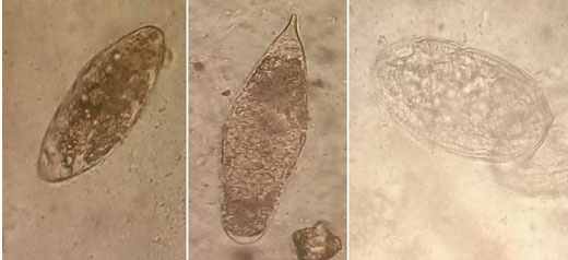
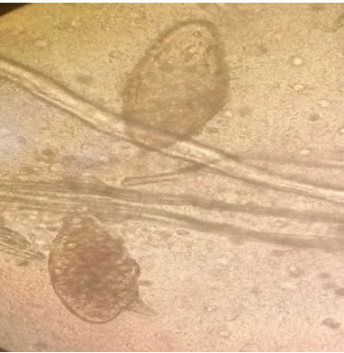
There were mixed Schistosoma infections in urine samples of the two cases. Urine sample of the 13 years old boy had terminal-spined eggs characteristic of S. haematobium, and other spineless oval eggs (Figure 1). While urine sample from the 14 years old boy contained both terminal-spined eggs that were characteristic of S. haematobium and sub-terminal-spined eggs suggestive of hybrid types as shown in Figure 2.
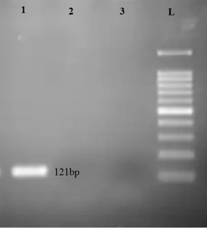
After subjecting the extracted genomic DNA from the mixed infections to PCR analysis, there was no amplification for the Dra 1 sequence of S. haematobium from both cases as shown on Lane 2 (from the 13 years old boy in Giwa LGA) and Lane 3 (from the 14 years old boy in Kaduna South LGA) in Figure 3. However, from the positive control, the 121bp Dra 1 tandem repeat sequence was amplified.
DISCUSSION
The urine samples were subjected to urinalysis (using SG11100-Uric 11V, Guilin Zhonghui Technology Co., Ltd, China), and both contained microhematuria, proteins, leukocytes, and ketones. Each urine sample was gently agitated to mix and divided into two equal volumes of 10 mL and subjected to centrifugation at 3000 revolutions per minute (rpm). One of the sediments was examined under 10× and 40× objectives of the light microscope for detection of eggs of Schistosoma. The other sediment was used for genomic DNA extraction using phenolchloroform method. The sediment was transferred onto a sterile test tube containing 200 µL nuclease-free-water and heated at 100°C for 10 minutes [6]. The mixture was vortexed briefly. A total of 250 µL of the mixture was added to 250 µL of buffer-saturated phenol/chloroform/isoamylalcohol (pH 7.5–7.8), and 25 µL of 3 M sodium acetate (pH 5) and vortexed for 1 minute. After 15 minutes incubation on ice, it was centrifuged for 10 minutes at 12,000 rpm at room temperature (26°C). The aqueous layer was transferred to a 1.5 mL Eppendorf tube containing 250 µL isopropanol and incubated for 5 minutes on ice. The mixture was centrifuged at 12,000 rpm at 4°C for 5 minutes. Supernatant was discarded and the pellet was re-suspended in 50 µL of 70% ethanol, vortexed and centrifuged at 13,000 rpm for 2 minutes at room temperature (30°C). The supernatant was discarded and the residual ethanol was eliminated by a quick centrifuge spin. The pellet was air-dried in a half-open tube for about 30 minutes and suspended in 20 µL of TE (10 mM TrisCl, 1 mM EDTA, pH 8), vortexed and stored at -20°C for polymerase chain reaction [7],[9]. The genomic DNA was electrophoresed and viewed under UV-light.
The Dra 1 primers were designed for amplification of 121bp tandem repeat sequence of S. haematobium, which was previously described by Hamburger et al. [10] and validated by Ibironke et al. [11] and used for S. haematobium DNA detection on filtered urine [12]. Dra 1 forward and reverse primers and other consumables were obtained from Inqaba Biotechnical Industries (Pty) Ltd., Pretoria, South Africa and prepared according to manufacturers’ instructions.
The polymerase chain reaction (PCR) was performed in a 25 µL reaction unit containing 12.5 µL of OneTaq Quick-Load 2X Mastermix with Standard Buffer (New England Biolab Inc.), 0.5 µL DRA1F (0.01 umol forward primer, 5'-GATCTCACCTATCAGACGAAAC-3'), 0.5 µL DRA1R (0.01 umol reverse primer, 5'-TCACAACGATACGACCAAC-3'), 5 µL S. haematobium genomic DNA (as template) and 6.5 µL nuclease-free water. The PCR cycling conditions included initial denaturation at 95°C for 5 minutes, followed by 33 cycles at 95°C for 30 seconds (denaturation), annealing at 47°C for 60 seconds and extension at 72°C for 1 minute and final extension at 72°C for 5 minutes. After which samples were kept on hold at 4°C. A positive control of extracted genomic DNA from urine with S. haematobium was used. The PCR products were visualized on 2% agarose gel.
Though the 14 years old boy did not present with any overt symptoms of the disease, urinalysis indicated evidence of microhematuria. Furthermore, both of them had proteins, leukocytes, and ketones in their urine samples. These urinary indices by application of urinalysis can serve as useful indicators of urinary schistosomiasis. Classically, urinary schistosomiasis is caused by S. haematobium, and recovered eggs were oval with terminal spines. The detection of elongated, nearly spindle-shaped eggs with terminal spines and elliptical spineless eggs are not commonly reported in cases of urinary schistosomiasis. Similarly, mixed terminal-spined eggs and sub-terminal-spined eggs are uncommon in Nigeria. These two cases of urinary schistosomiasis are rare occurrences of mixed infections. Majority of prevalent Schistosoma mixed infections are between S. haematobium and S. mansoni, which are human parasites [13],[14]. Study by Ojo et al. [15] in South West Nigeria suggested the possibility of interspecific interactions between S. haematobium and S. mansoni, as distinctive eggs of the two species were found regardless of predilection sites. Typically, terminal-spined eggs and lateral-spined eggs are discharged through urine and stool respectively, and common in co-infections. Both eggs with bump-like inconspicuous sub-terminal spines (Figure 1) and the elliptical egg with sub-terminal spines are much more of morphotypes of S. bovis. Such variations in egg morphotypes had been previously reported in Europe by Kincaid-Smith et al. [16].
Abnormal routing of predilection sites by Schistosoma species had been reported [17]. During heavy Schistosoma infections, the eggs can be carried to any location in the body, capable of causing tissue damage [18]. Hybridization had also been reported among mammalian schistosomes that are phylogenetically related in regions where distinctive snail hosts co-exist [7],[19]. Cattle rearing methods like transhumance and nomadism are responsible for movement of domestic livestock, permitting animal contact with multiple potential sites for schistosomiasis transmission [20].
Hybrids of Schistosoma species that infect man exist. Those species that infect animals and man can also hybridize [21]. A major concern is on the epidemiology and zoonotic transmission of human–animal schistosome hybrids [20]. Hybrid species of S. haematobium (which lives mainly in the venous plexus of human bladder) and S. bovis (mainly in mesenteric veins of animal intestine) had been reported in humans [22] in some West African countries, especially in Senegal [23],[24], Mali [25], and recently becoming endemic in South Europe [16],[26]. There is no comprehensive information on Schistosoma species genomic diversity in Nigeria yet, compared with data from neighboring countries.
Giwa and Kaduna South LGAs are cattle-breeding areas where these rare cases of mixed urinary schistosomiasis were found in Northern Nigeria. In cattle breeding areas, both humans and the cattle share the same rivers. Bulinus snails serve as intermediate host for both S. haematobium and S. bovis. Within the snail or human host, hybridization can occur. Hybrid species of Schistosoma discharge morphologically highly polymorphic eggs [16].
There was no amplification of Dra 1 tandem sequence of S. haematobium in both cases of mixed urinary Schistosoma infections. This directly implied that the species in these cases where not classically S. haematobium, but probably some hybrid species causing urinary schistosomiasis, and may be circulating in Nigeria. Zoonotic schistosomiasis can undermine control efforts on schistosomiasis, especially in Africa.
CONCLUSION
In this report, the two cases of mixed urinary schistosomiasis from Kaduna State in Nigeria had microhematuria, proteinuria, leukocyturia, and ketonuria as detected by urinalysis. Such indices can be of diagnostic importance in urinary schistosomiasis. Microscopic examination of the cases revealed rare occurrences of mixed infections of egg morphotypes of Schistosoma species (other than S. haematobium), suggestive of hybridization phenomenon.
REFERENCE
1.
Schistosomiasis. [Available at: https://www.who.int/news-room/fact-sheets/detail/schistosomiasis]

2.
Bishop HG, Inabo HI, Ella EE. Prevalence and intensity of urinary schistosomiasis and their effects on packed cell volume of pupils in Jaba LGA, Nigeria. Edorium J Microbiol 2016;2:13–26.
3.
Bishop HG. Menace of schistosomiasis: Its true neglected nature in Nigeria. MOJ Public Health 2017;6(5):421–6. [CrossRef]
4.
Parasites - Schistosomiasis. [Available at: https://www.cdc.gov/parasites/schistosomiasis/index.html]
5.
Akinwale OP, Hock TT, Chia-Kwung F, et al. Differentiating Schistosoma haematobium from Schistosoma magrebowiei and other closely related schistosomes by polymerase chain reaction amplification of a species specific mitochondrial gene. Trop Parasitol 2014;4(1):38–42. [CrossRef]
[Pubmed]
6.
Hessler MJ, Cyrs A, Krenzke SC, et al. Detection of duo-schistosome infection from filtered urine samples from school children in Zambia after MDA. PLoS One 2017;12(12):e0189400. [CrossRef]
[Pubmed]
7.
Abbasi I, Webster BL, King CH, Rollinson D, Hamburger J. The substructure of three repetitive DNA regions of Schistosoma haematobium group species as a potential marker for species recognition and interbreeding detection. Parasit Vectors 2017;10(1):364. [CrossRef]
[Pubmed]
8.
Mogaji HO, Omitola OO, Bayegun AA, Ekpo UF, Taylor-Robinson AW. Livestock reservoir hosts: An obscured threat to control of human schistosomiasis in Nigeria. Zoonotic Dis 2023;3(1):52–67. [CrossRef]
9.
Sarhan RM, Kamel HH, Saad GA, Ahmed OA. Evaluation of three extraction methods for molecular detection of Schistosoma mansoni infection in human urine and serum samples. J Parasit Dis 2015;39(3):499–507. [CrossRef]
[Pubmed]
10.
Hamburger J, He-Na, Abbasi I, Ramzy RM, Jourdane J, Ruppel A. Polymerase chain reaction assay based on a highly repeated sequence of Schistosoma haematobium: A potential tool for monitoring schistosome-infested water. Am J Trop Med Hyg 2001;65(6):907–11. [CrossRef]
[Pubmed]
11.
Ibironke OA, Phillips AE, Garba A, Lamine SM, Shiff C. Diagnosis of Schistosoma haematobium by detection of specific DNA fragments from filtered urine samples. Am J Trop Med Hyg 2011;84(6):998–1001. [CrossRef]
[Pubmed]
12.
Ibironke O, Koukounari A, Asaolu S, Moustaki I, Shiff C. Validation of a new test for Schistosoma haematobium based on detection of Dra1 DNA fragments in urine: Evaluation through latent class analysis. PLoS Negl Trop Dis 2012;6(1):e1464. [CrossRef]
[Pubmed]
13.
Meurs L, Mbow M, Vereecken K, Menten J, Mboup S, Polman K. Epidemiology of mixed Schistosoma mansoni and Schistosoma haematobium infections in northern Senegal. Int J Parasitol 2012;42(3):305–11. [CrossRef]
[Pubmed]
14.
Lodh N, Naples JM, Bosompem KM, Quartey J, Shiff CJ. Detection of parasite-specific DNA in urine sediment obtained by filtration differentiates between single and mixed infections of Schistosoma mansoni and S. haematobium from endemic areas in Ghana. PLoS One 2014;9(3):e91144. [CrossRef]
[Pubmed]
15.
Ojo JA, Adedokun SA, Akindele AA, et al. Prevalence of urogenital and intestinal schistosomiasis among school children in South-west Nigeria. PLoS Negl Trop Dis 2021;15(7):e0009628. [CrossRef]
[Pubmed]
16.
Kincaid-Smith J, Tracey A, de Carvalho Augusto R, et al. Morphological and genomic characterisation of the Schistosoma hybrid infecting humans in Europe reveals admixture between Schistosoma haematobium and Schistosoma bovis. PLoS Negl Trop Dis 2021;15(12):e0010062. [CrossRef]
[Pubmed]
17.
Atanda AT, Mohammad MS, Atallah L. Cutaneous schistosomiasis: Case report and literature review. Annals of Nigerian Medicine 2012;6(2):98–100.
18.
Cheesbrough M. District Laboratory Practice in Tropical Countries, Part I. 2ed. Cambridge, UK: Cambridge University Press; 2009.
19.
Schistosomiasis: Epidemiology and clinical manifestations. [Available at: https://www.uptodate.com/contents/schistosomiasis-epidemiology-and-clinical-manifestations]
20.
Djuikwo-Teukeng FF, Kouam Simo A, Allienne JF, et al. Population genetic structure of Schistosoma bovis in Cameroon. Parasit Vectors 2019;12(1):56. [CrossRef]
[Pubmed]
21.
Leger E, Webster JP. Hybridizations within the Genus Schistosoma: Implications for evolution, epidemiology and control. Parasitology 2017;144(1):65–80. [CrossRef]
[Pubmed]
22.
Calvo-Cano A, Cnops L, Huyse T, et al. A case of urogenital human schistosomiasis from a non-endemic area. PLoS Negl Trop Dis 2015;9(11):e0004053.
[Pubmed]
23.
Huyse T, Webster BL, Geldof S, et al. Bidirectional introgressive hybridization between a cattle and human schistosome species. PLoS Pathog 2009;5(9):e1000571. [CrossRef]
[Pubmed]
24.
Huyse T, Van den Broeck F, Hellemans B, Volckaert FAM, Polman K. Hybridisation between the two major African schistosome species of humans. Int J Parasitol 2013;43(8):687–9. [CrossRef]
[Pubmed]
25.
Soentjens P, Cnops L, Huyse T, et al. Diagnosis and clinical management of Schistosoma haematobium-Schistosoma bovis hybrid infection in a cluster of travelers returning from Mali. Clin Infect Dis 2016;63(12):1626–9. [CrossRef]
[Pubmed]
26.
Boissier J, Grech-Angelini S, Webster BL, et al. Outbreak of urogenital schistosomiasis in Corsica (France): An epidemiological case study. Lancet Infect Dis 2016;16(8):967–9. [CrossRef]
[Pubmed]
SUPPORTING INFORMATION
Acknowledgement
We express our appreciation to the two consented adolescents, who were the cases in this report. We equally thank Dr. Y.Y. Pala for his kind contribution.
Author ContributionsHenry Gabriel Bishop - Conception of the work, Design of the work, Acquisition of data, Analysis of data, Drafting the work, Final approval of the version to be published, Agree to be accountable for all aspects of the work in ensuring that questions related to the accuracy or integrity of any part of the work are appropriately investigated and resolved.
Helen Ileigo Inabo - Conception of the work, Design of the work, Revising the work critically for important intellectual content, Final approval of the version to be published, Agree to be accountable for all aspects of the work in ensuring that questions related to the accuracy or integrity of any part of the work are appropriately investigated and resolved.
Elijah Ekah Ella - Analysis of data, Revising the work critically for important intellectual content, Final approval of the version to be published, Agree to be accountable for all aspects of the work in ensuring that questions related to the accuracy or integrity of any part of the work are appropriately investigated and resolved.
Mohammed Bello - Revising the work critically for important intellectual content, Final approval of the version to be published, Agree to be accountable for all aspects of the work in ensuring that questions related to the accuracy or integrity of any part of the work are appropriately investigated and resolved.
Guarantor of SubmissionThe corresponding author is the guarantor of submission.
Source of SupportNone
Consent StatementWritten informed consent was obtained from the patient for publication of this article.
Data AvailabilityAll relevant data are within the paper and its Supporting Information files.
Conflict of InterestAuthors declare no conflict of interest.
Copyright© 2023 Henry Gabriel Bishop et al. This article is distributed under the terms of Creative Commons Attribution License which permits unrestricted use, distribution and reproduction in any medium provided the original author(s) and original publisher are properly credited. Please see the copyright policy on the journal website for more information.