 |
|
Case Report
| ||||||
| Alcohol and acute pancreatitis contributing to central pontine myelinolysis | ||||||
| Sterling Farrer | ||||||
|
Western University of Health Sciences, Lebanon
| ||||||
| ||||||
|
[HTML Abstract]
[PDF Full Text]
[Print This Article] [Similar articles in PubMed] [Similar articles in Google Scholar] |


| How to cite this article |
| Farrer S. Alcohol and acute pancreatitis contributing to central pontine myelinolysis. Case Rep Int 2018;7:100048Z06SF2018. |
|
ABSTRACT
| ||||||||||||||||||||||||||||||||||||||||||
|
Introduction: Central pontine myelinolysis (CPM) is a well-recognized syndrome that is related to various conditions such as rapid correction of hyponatremia and chronic alcoholism. Case Report: We report a case of a recently discharged patient with dysarthria, vertigo, and progressive gait changes, with radiological evidence of CPM, but without the expected rapid correction of hyponatremia seen in other patients that developed CPM. Conclusion: CPM is rare without severe hyponatremia of 120 meq/L or less, but there are other factors that contribute to the development of CPM, that may be unrelated to hyponatremia. Severe alcohol abuse, acute pancreatitis, and subsequent poor nutritional intake were likely the major factors in inducing osmotic injury in this case, which lead to CPM. Keywords: Alcoholism, Central pontine myelinolysis, Hyponatremia, Osmotic demyelination syndrome, Pancreatitis | ||||||||||||||||||||||||||||||||||||||||||
|
INTRODUCTION
| ||||||||||||||||||||||||||||||||||||||||||
|
Central Pontine myelinolysis (CPM), also known as osmotic demyelination syndrome, is defined as a neurological disorder caused by damage of the myelin sheath of the nerve cells in the pons [1]. It was first described in 1959 as a disease affecting alcoholics and the malnourished [2]. The cause was not known then but the authors suspected either a toxin or a nutritional deficiency. Many studies have implicated the rapid correction of hyponatremia as the major factor associated with CPM, due to exposing the pontine glia and extrapontine glia to osmotic stress [3]. CPM presents most commonly as a complication of treatment of patients with profound hyponatremia [4]. There are other case reports of patients developing CPM that are not related to profound hyponatremia or the rapid correction of hyponatremia. These case reports have found the cause to be associated with alcohol withdrawal, hypokalemia, anorexia nervosa when feeding is started, patients undergoing dialysis, and burns victims [5], [6], [7]. We present a case of CPM associated with alcohol abuse and acute pancreatitis without profound hyponatremia, or rapid correction of hyponatremia. | ||||||||||||||||||||||||||||||||||||||||||
|
CASE REPORT
| ||||||||||||||||||||||||||||||||||||||||||
|
A 52-year-old female with a history of alcoholic pancreatitis and atrial fibrillation presented with gradually worsening weakness and gait changes for eight days. She was admitted two weeks previously for alcohol induced pancreatitis. She initially hid the information of her heavy alcohol drinking which delayed the diagnosis process, and she had a hospital stay of 10 days. During her last admission, she initially had some hyponatremia with sodium level 122, and was corrected slowly to 132 in 3 days. She was discharged eight days later in good condition, with no pain or weakness. Since that time she reported gradually developing bilateral leg weakness and gait changes. She reported multiple falls. On one occasion she was walking into her home, lost balance, and stumbled into a bush. She stated her legs felt like noodles and that she felt like the ground was spinning when she started to fall down. She also had progressive speech changes since being discharged. The morning before coming back to the emergency department, she was getting juice out of the fridge when the room started to spin. Her legs became very weak and she stumbled to the floor. After 30 minutes of lying on the floor, she was able to crawl to her couch and call for help. On physical examination, there we no focal deficits, and she showed normal sensation, muscle strength and reflexes. Labs were obtained on admission and her complete labs were: Basic Metabolic Panel, Sodium 134 mmol/L, Potassium 3.9 mmol/L, Chloride 97 mmol/L, Bicarbonate 23.6 mmol/L, Glucose 201 mg/dL, Creatinine 0.7 mg/dL, Calcium 9.2 mg/dL, Anion Gap 13, BUN/Creatinine Ratio 13.4 Complete Blood Count with Differential:
An MRI was acquired and the radiologist reported, “Pontine signal abnormality with mildly expansive appearance. Evidence of possible bilateral pontine myelinolysis.” Admission labs showed the patient has no electrolyte abnormalities. The patient’s blood sodium level at the time was 131 meq/L. The suggested emergent treatment for CPM is to relower the sodium level to a value that is less than 16 meq/L above the initial serumsodium. Since review of previous admission records showed a slowly corrected hyponatremia, that raised sodium levels only 10 meq/L over three days, this was not necessary. Patient was treated with supportive therapy, which included thiamine, vitamin B12, and folic acid. Supportive therapy also included working with physical therapy and speech therapy. Patient’s neurologic deficits slightly improved over her four day admission, and she was discharged to a rehabilitation center. | ||||||||||||||||||||||||||||||||||||||||||
DISCUSSION | ||||||||||||||||||||||||||||||||||||||||||
|
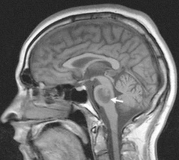
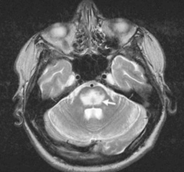
Central pontine myelinolysis (CPM) is a neurological disorder caused by damage of the myelin sheath of the nerves surrounding the pons. This lesion can be identified in both T1 and T2 signal intensity MRI, shown above in Figures 1 and 2 [8]. It is characterized by acute paralysis, gait changes, dysarthria, and other neurologic symptoms. The clinical presentation in this case was typical for CPM. Considering the acute and progressive nature of the patient’s lower extremity weakness, directly following a ten-day hospital admission, one would suspect an iatrogenic, treatment induced cause. The most common cause of CPM is overly rapid correction of low blood sodium levels. A closer look at the patient’s lab history from their previous history showed that the patient was hyponatremic upon admission, with a blood sodium level of 122 meq/L (normal range 125–135). The patient’s hyponatremia was corrected over a span of three days, eventually reaching 132 meq/L on the third day. The majority of ODS cases occur in patients whose sodium concentrations at presentation are ≤105 meq/L, and the vast majority of reported cases arise in patients who present with a serum sodium of 120 meq/L or less [9]. When the initial serum sodium is greater than 120 meq/L, ODS is rare and is typically limited to patients with severe liver disease or patients with diabetes insipidus who have developed a moderate degree of hyponatremia as a complication of desmopressin therapy and then have the desmopressin discontinued [10]. Our patient had neither of these clinical scenarios, and had a serum sodium of over 120 meq/L. Studies in experimental animals have shown that osmotic demyelination is caused by correction of hyponatremia and not by hyponatremia itself, and that more rapid rates of correction increase the likelihood and severity of brain lesions [11]. The threshold rate of correction, above which brain lesions may develop, is approximately 15 meq/L per day in dogs and 16 meq/L in rats; the incidence of lesions progressively increases with larger magnitudes of daily correction [11]. Observational studies in humans are consistent with experimental studies in animals, showing that more rapid rates of correction of chronic hyponatremia are associated with osmotic demyelination [8]. With our patient’s sodium rate of 122 being corrected to 132 over a span of 3 days, it does not seem that rapid correction of hyponatremia is likely to be the cause of her CPM. Apart from rapid correction of hyponatremia, there are case reports of central pontine myelinolysis in association with hypokalemia, anorexia nervosa when feeding is started, and chronic alcoholism [12]. In these instances, occurrence may be entirely unrelated to hyponatremia or rapid correction of hyponatremia. In our patient’s case, a combination of these factors played a role in the development of CPM. Before the patient was admitted for pancreatitis, she reported heavy alcohol abuse due to depression, drinking multiple bottles of wine a day. The direct toxicity of alcohol induces osmotic injury to the vascular endothelial cells and this causes the release of myelinotoxic factors, the production of vasogenic edema. This then causes separation of the axon from its myelin sheath with resultant injury to the oligodendrocytes [5]. Recent acute pancreatitis may have also played a role in the development of CPM. A previous study suggested that acute pancreatitis may result in abnormalities of astrocyte metabolism with a reduced ability to generate new intracellular anions in response to anion change [13]. Demyelination may then occur with changes in serum osmolality. In this case, even a moderate serum osmolality change, such as the correction of sodium levels from 122 to 132, could result in osmotic endothelial injury and opening of the blood brain barrier. | ||||||||||||||||||||||||||||||||||||||||||
| ||||||||||||||||||||||||||||||||||||||||||
| ||||||||||||||||||||||||||||||||||||||||||
|
CONCLUSION
| ||||||||||||||||||||||||||||||||||||||||||
|
In conclusion, although CPM is rare without severe hyponatremia of 120 meq/L or less, there are other factors that contribute to the development of CPM, that may be unrelated to hyponatremia. In this patient’s case, their borderline low sodium levels at presentation did not likely cause CPM. The severe alcohol abuse, acute pancreatitis, and subsequent poor nutritional intake were likely the major factors in inducing osmotic injury leading to injury to the oligodendrocytes, with resultant CPM. | ||||||||||||||||||||||||||||||||||||||||||
|
REFERENCES
| ||||||||||||||||||||||||||||||||||||||||||
| ||||||||||||||||||||||||||||||||||||||||||

|
[HTML Abstract]
[PDF Full Text]
|
|
Author Contributions
Sterling Farrer – Substantial contributions to conception and design, Acquisition of data, Analysis and interpretation of data, Drafting the article, Revising it critically for important intellectual content, Final approval of the version to be published |
|
Guarantor of Submission
The corresponding author is the guarantor of submission. |
|
Source of Support
None |
|
Consent Statement
Written informed consent was obtained from the patient for publication of this case report. |
|
Conflict of Interest
Author declares no conflict of interest. |
|
Copyright
© 2018 Sterling Farrer. This article is distributed under the terms of Creative Commons Attribution License which permits unrestricted use, distribution and reproduction in any medium provided the original author(s) and original publisher are properly credited. Please see the copyright policy on the journal website for more information. |
|
|